
Keratoacanthoma is a benign tumor that develops from sebaceous – hair follicles. The tumor grows very quickly, in the absence of treatment, the keratoacanthoma spontaneously regresses, leaving behind a very ugly scar. At risk for the occurrence of keratoacanthoma are people with fair skin, living in areas with intense sun exposure or in areas of industrial production. The Japanese and black Africans are not prone to this disease. Keratoacanthoma affects middle-aged and elderly people, more often men. Learn more about the manifestations of keratoacanthoma.
What are the causes and mechanism of development of keratoacanthoma?
Because keratoacanthoma appears when exposed to sunlight, swelling appears on exposed skin.
It has been established that chemical carcinogens such as mineral oils and tar provoke the development of keratoacanthoma. Mechanical trauma is also a predisposing factor.
Keratoacanthoma may appear on areas of the skin already affected by eczema or psoriasis. Read more about the symptoms of keratoacanthoma further on estet-portal.com. The viral nature of tumors is disputed, despite the fact that virus-like particles have been found in keratoacanthoma. After the phase of intensive growth of keratoacanthoma, the phase of rest, spontaneous involution and healing begins. This cyclicity is associated with the cycle of development of hair follicles. Most researchers dispute the involvement of immune mechanisms in the process.
Symptoms and clinical manifestations of the development of keratoacanthoma
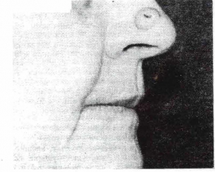
Keratoacanthoma is localized in most cases on the dorsal surface of the hands and on the middle zone of the face, however, it can appear on any part of the skin. The tumor can grow under the nail, causing pain in the underlying tissues. On the mucous membranes of keratoacanthoma appear rarely. Initially, the tumor looks like a small semicircular nodule, a bit like molluscum contagiosum.
Keratoacanthoma characteristic:
- Keratoacanthoma is flesh or red.
- The focus is growing rapidly and in a month – the second reaches sizes up to 3 cm in diameter.
- After that comes the stage of stabilization, then - spontaneous involution for 4-6 months.
- The tumor gradually grows and takes the form of a kidney, which has a characteristic crater-like depression in the center. As a rule, it is filled with horny masses, sometimes covered with a layer of skin.
- Over time, these masses in the center are rejected, leaving behind a depressed or wrinkled scar.
Key clinical course of keratoacanthoma
One variant of keratoacanthoma is a giant keratoacanthoma, the diameter of which can exceed 5 cm. It can grow in depth and destroy underlying tissues.
Another rare variant is centrifugal keratoacanthoma, which has rapid growth in the periphery with simultaneous involution and atrophy in the center. Spontaneous resolution is possible, but growth may continue for several years. This type of keratoacanthoma can appear on the trunk, face or limbs, reaching a size of more than 20 cm.
Multiple keratoacanthoma is quite rare. The clinical picture is usually sufficient to establish the diagnosis of keratoacanthoma.
Keratoacanthoma can be treated by various methods, including surgical excision, radiation therapy, Mohs surgical technique, curettage, electrosurgery, argon laser therapy, cryotherapy, corticosteroids, systemic retinoids, cytostatics, and photodynamic therapy.
The choice of treatment for keratoacanthoma depends on the location and size of the tumor.
Share:


Add a comment