
Kaposi's sarcoma (Kaposi's angiosarcoma, Kaposi's angioreticulosis, multiple hemorrhagic sarcoma) – a malignant multifocal disease, probably of a tumor nature, arising in the dermis and originating from the blood and lymphatic vessels, the foci are localized more often in the distal parts of the upper and lower extremities. In the initial stage, capillary-type vessels with wide gaps grow in the dermis. Endothelial cells are enlarged, elongated, nuclei of irregular shape, hyperchromic. Vascular proliferates are surrounded by edematous connective tissue infiltrated by lymphocytes and plasma cells. Expanded bloodless capillary spaces are separated by layers of collagen fibers.
Clinical manifestations and stages of Kaposi's sarcoma
Distinguish between classic (idiopathic) Kaposi's sarcoma and AIDS-associated (epidemic) sarcoma. Classical Kaposi's sarcoma proceeds for a long time, rarely generalizes with damage to the internal organs. AIDS – associated – may be the only manifestation of HIV infection, notes estet-portal.com. First, small bright pink or blue spots appear, & nbsp; resembling hematoma. Perhaps the appearance of red compacted plaques. In the future, dark red, purple or almost black painful nodes, ulcers appear.
Kaposi's sarcoma can develop with immunosuppressants after organ transplantation or with long-term use of drugs for chronic dermatoses.
At first, manifestations on the skin are limited, over time, the disease becomes aggressive. With a decrease in the dose of immunosuppressants, skin and visceral manifestations regress in 24-80% of cases.
The following stages of Kaposi's sarcoma are distinguished according to the clinical picture:
- Spotted stage – characterized by individual reddish-cyanotic or reddish-brown spots of irregular shape up to 1-5 cm in diameter with a smooth surface.
- Papular stage - in which the tumor has a spherical or hemispherical shape, diameter 2mm – 1 cm, color from pink to reddish-bluish with a brown tint, densely elastic consistency.
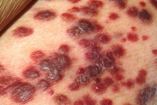
- Tumor stage – it is characterized by multiple nodes of red-cyanotic or cyanotic-brown color, soft or densely elastic on palpation, with a diameter of 1-5 cm. The nodes, increasing in size, merge into bumpy painful formations, eventually covered with ulcers with copious secretions and an unpleasant odor. Ulcers invade the subcutaneous fat, often bleed. The affected limbs swell, become covered with a hemorrhagic rash, rarely there are blisters with bloody contents. With Kaposi's sarcoma often the lymph nodes increase, in 10-12% of cases the mucous membranes of the mouth are affected, less often the genitals.
Aspects of diagnosis and treatment of Kaposi's sarcoma
Diagnosis of Kaposi's sarcoma is based on the clinical presentation and histological findings. Patients with Kaposi's sarcoma must be examined for HIV infection.
Kaposi's sarcoma should be differentiated from Kaposi's pseudosarcoma, hemangioma, pyogenic granuloma, well-differentiated angiosarcoma.
Pathohistological examination of the biopsy of the affected skin reveals chaotic incomplete angiogenesis, proliferation of spindle cells, proliferation of granulation tissue of varying degrees of maturity, infiltrated by immunocompetent cells (lymphocytes, plasma cells and macrophages). In the initial stage, capillary-type vessels with wide lumens grow in the upper part of the dermis.
Treatment of Kaposi's sarcoma is not always effective, existing means (applications of 30% prospidin ointment, radiation methods, cryotherapy,chemotherapy, applications of dinitrochlorobenzene, injections of interferon-alpha) cause only temporary remission. For large or painful lesions, x-rays are used.
A noticeable therapeutic effect during the course of the disease occurs after the systemic use of prospidin and cytostatics.
Sick AIDS – Associated Kaposi's sarcoma is also prescribed drugs used to treat HIV – infected.
Share:


Add a comment