


Bullous pemphigoid – this is a chronic autoimmune disease from the group of vesicular dermatoses, which is characterized by a significant degree of prevalence and often – death.
Another name for the disease – non-acantholytic pemphigus: the appearance of subepidermal blisters is not accompanied by signs of acantholysis. Lever's pemphigoid more often affects children and the elderly over 60 years of age.
The disease is acquired: bullous pemphigoid is often associated with other autoimmune diseases.
On estet-portal.com read about the pathogenesis, main clinical criteria and methods for diagnosing bullous pemphigoid, as well as the relationship of dermatosis with other immune diseases.
Features of the course of bullous pemphigoid and clinical symptoms of the disease
Lever's pemphigoid often begins with symmetrical lesions resembling erythema multiforme.
There are two periods of the course of the disease – prodromal and bullous.
During the prodromal period, it can be difficult to diagnose the disease: the only clinical symptom at this time may be only itching for several months.
The bullous period is characterized by the appearance of a rash in the form of intense subepidermal blisters with serous or serous-hemorrhagic contents.
Follow us on Facebook
Favorite localization of – trunk, limbs, as well as the area of \u200b\u200bnatural folds. Further bubbles are opened with formation of painful erosions and in the subsequent – flat crusts on the skin.
Mucosal lesions occur as a manifestation of the vesicular form of the disease: mostly bubbles appear on the hard palate and cause discomfort in patients.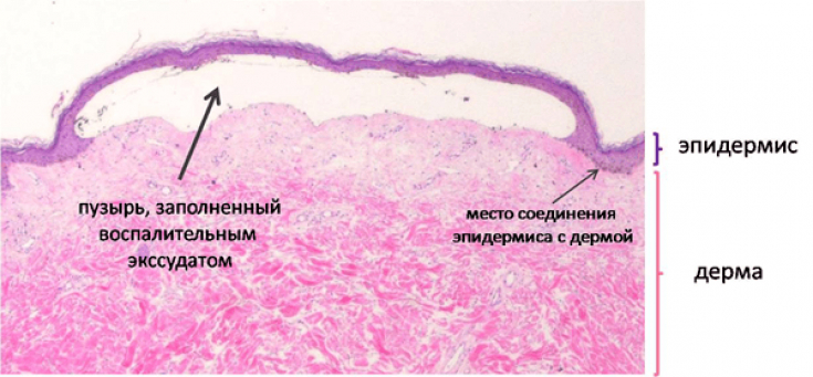
Pathogenesis of acquired pemphigoid: mechanisms and trigger factors
The etiology of bullous pemphigoid has not been fully understood to date. It is known that autoimmune reactions play an important role in the development of the disease: the interaction of autoantibodies with surface antigens with basal layer antigens leads to the activation of complement and inflammatory mediators.
The surface antigens BPAG1 and BPG2 are proteins within the hemidesmosomes – structures of the intercellular space of the epidermis.
This releases proteases that cleave desmosomal proteins and lead to blister formation.
But in order for such a process to develop, it is necessary to influence trigger factors, such as:
• chronic dermatoses;
• drugs (NSAIDs, cephalosporins, penicillins, ACE inhibitors);
• viral infections (cytomegalovirus, hepatitis B and C viruses, Epstein-Barr virus,);
• mechanical and physical influences (ultraviolet irradiation, thermal burns, radiation therapy). therefore, older people with a weakened immune system and chronic diseases are most often ill.
You may also be interested in:
Dermatosis blisteris: what is important to remember
Diseases associated with bullous pemphigoid

Pemphigoid can also occur in people with neurological diseases such as Parkinson's disease, dementia, bipolar disorders, paralysis, multiple sclerosis.
An important role in the development of the disease is played by other autoimmune systemic diseases, such as SLE, dermatomyositis, rheumatoid arthritis, Hashimoto's thyroiditis and others.
Neacantholytic pemphigus often manifests itself in patients with oncopathologies: malignant neoplasms or lymphoproliferative diseases.
How dangerous is Duhring's dermatitis herpetiformis
Diagnosis of pemphigoid: laboratory, serological and differential
The diagnosis of bullous pemphigoid is based on the characteristic clinical presentation, laboratory and serological studies.
Main diagnostic methods include:1. histological analysis (spongiosis, increased content of eosinophils - more than 20-30%);
2. RIF (IgG along the basement membrane);
3. ELISA (circulating IgG antibodies to BPAG1 or BPAG2);
4. immunoblotting or immunoprecipitation.
Bullous pemphigoid must be differentiated from diseases such as dermatitis herpetiformis, allergic contact dermatitis, congenital epidermolysis bullosa, urticaria, gestational pemphigoid, Stevens-Johnson syndrome, bullous mastocytosis and other blistering forms of dermatoses.
Thank you for staying with estet-portal.com. Read other interesting articles in the "Dermatology" section. You may also be interested in:
Polymophoric skin rash: how to recognize erythema multiforme