
There are many reasons why patients seek medical help from a urologist, but most urological pathologies do not require immediate medical attention. Urolithiasis is precisely that urological disease that is asymptomatic for a long time, and manifests itself spontaneously and acutely in the form of renal colic. Many different factors influence the formation of kidney stones, and knowledge about the types and sizes of stones plays an important role in the choice of treatment tactics. So, how are kidney and ureter stones formed and what are distinguished.
What factors influence the formation of kidney stones
Many different factors influence the development of urolithiasis. In order for a stone to form in the kidney, two main conditions are necessary: the etiological factor and the conditions predisposing to stone formation. The etiological factor, in fact, is the physicochemical processes of violation of crystallization and colloidal equilibrium. In this case, urine should contain a supersaturated solution of salts, from which the stone is formed. At the same time, the content in the urine of substances that inhibit the formation of crystals should be reduced. The main factors predisposing to the formation of kidney stones are concomitant diseases such as tubulopathies and chronic pyelonephritis, lifestyle, diet and drinking regimen.
Classification of kidney stones by their chemical composition
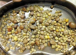
The chemical composition of kidney stones depends primarily on the general condition of the body and local factors. Each stone has an organic matrix that is coated with calcium salts. There are these types of kidney stones:
- calcium oxalate stones – consist of calcium salts of oxalic acid, their crystals are thorn-shaped, colored in colors from dark brown to black;
- phosphate and phosphate-magnesium stones – contain calcium salts of phosphoric acid, they are round, gray in color, the stones are quite soft and loose;
- carbonate stones – consist of calcium salts of carbonic acid, have a homogeneous composition, white color, rather friable;
- urate stones – stones of uric acid and its salts, hard, even, brick-yellow;
- cystine stones – consist of the sulfur compound of the amino acid cystine, yellow in color, round in shape, have a smooth surface and a soft texture;
- cholesterol stones – composed of cholesterol, soft, brittle stones of black color;
- protein stones – consist of fibrin with an admixture of salts and bacteria, white, soft and flat.
Coral stones – a special kind of kidney stones
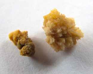
Special attention should be paid to staghorn kidney stones, the special form of which determines the clinical course of this type of urolithiasis. Coral stones are composed of ammonium, magnesium phosphate and calcium phosphate. Such stones are localized in the renal pelvis and cups, and as they grow, they can spread to the entire cup system. Coral-like stones, like a cast, repeat the shape of the pyelocaliceal system of the kidney. In the process of growth of coral stones, three main degrees are distinguished:
- In the first degree, a kidney stone is almost completely located in the renal pelvis, but has peculiar processes towards the cups of the kidney.
- At the second degree, the processes of staghorn stone fill all the large cups of the kidney.
- At the third degree, a staghorn stone completely fills the renal pelvis and calyces, forming branches at the level of the small calyxes.
At what levels can kidney stones cause obstruction
Kidney stones can be single or multiple, which is of fundamental importance for the choice of treatment tactics. During the diagnosis, it is important to clarify the shape, size and localization of stones, since the place where stones can cause obstruction largely determines the clinical picture and the subsequent course of the pathological process. There are three main risk areas where a stone can cause an obstruction:
- pelvic-ureteral segment – in this area, the wide pelvis narrows to 2-3 mm. and passes into the ureter, having a diameter of up to 19 mm.;
- the area where the ureter crosses the upper edge of the entrance to the pelvis – when crossing with the iliac vessels, the diameter of the ureter narrows to 3-4 mm.;
- vesicoureteral segment – the ureter in this area also narrows to 2-4 mm.
Share:


Add a comment