
Laser – abbreviation of English origin: LASER – "Light Amplification by Stimulated Emission of Radiation", which means "light amplification by stimulated emission". In other words, the laser – it is a device capable of producing a very powerful beam of monochromatic light. Since the laser beam – it is just a stream of light (albeit with some features), then later in this article it is called a beam of light.
Light – it is an electromagnetic (EM) wave that propagates in space at a tremendous speed (in a vacuum: c = 300,000 km/s). Unlike acoustic and mechanical waves, EM waves include two components – electrical and magnetic, - harmonic oscillations of which occur in mutually perpendicular directions. On the other hand, we can assume that the light flux consists of special particles (photons), the energy of which is related to the frequency of light ( E=hw, h – Dirac constant), and the number – with beam intensity.

Vladimir Aleksandrovich Tsepkolenko
MD, Professor, Honored Doctor of Ukraine,
President of the Ukrainian Society of Aesthetic
Medicine, General Director of the Ukrainian
Institute of Plastic Surgery
and Aesthetic Medicine "Virtus"
Light and its propagation in a homogeneous medium
The main characteristic of light – its frequency w, which determines the transferred energy. Light with different frequencies is perceived as different colors. For example, the frequency of red is less than the frequency of yellow, and yellow – less than blue. All possible frequencies of light are united by the term spectrum.
In visible light there is not one, but an infinite number of waves with different frequencies, which enter it in different proportions. Such a set of frequencies is called the spectral composition of light (in everyday life it is called color). If the stream of light "contains" waves of only one frequency, then it is called monochromatic (however, ideally monochromatic light cannot exist).
The second important characteristic of the luminous flux is its intensity I, which is directly related to the energy transferred in one second.
The concept of frequency is inconvenient in that its numerical values are unusually large for us, so another physical quantity is more often used – wavelength λ:

The higher the frequency of light, the shorter its wavelength. When light passes from one medium to another, its wavelength changes, but its frequency remains unchanged. Usually this fact is omitted, mentioning the wavelength not in the medium under consideration, but corresponding to it in vacuum.
Visible range radiation is called EM waves perceived by the human eye, the wavelengths of which lie in the range from 400 to 760 nm (Table 1).

Infrared refers to radiation with wavelengths greater than 760 nm (red), it is no longer visible, but we feel it as heat coming from any heated body.
Ultraviolet, on the contrary, refers to radiation in the range of 6-400 nm.
Reflection and refraction of light at the interface
In a homogeneous medium, a beam of light always forms a straight line. Light does not change direction on its own, but if an obstacle is encountered in the path of the beam in the form of a dust particle, droplet, or the boundary of another medium, it can change the direction of its movement. Such processes are called scattering or refraction.
Each medium (whether liquid, gas or transparent solid) is characterized by some quantity – refractive index n. The greater the difference between the refractive indices, the more the light is refracted. It is worth noting that light incident at right angles to the interface is not refracted, but continues to move in a straight line.
Another effect that occurs when light passes through an interface is its reflection from that interface. Reflection occurs almost always, and it is the greater, the smaller the angle between the beam and the interface between the media (the beam, as it were, ricochets from it). If light enters an inhomogeneous medium, then it is scattered. When scattered, part of the light is almost always "reflected", changing the direction of movement to the opposite.
Scattering and reflection effects, as a rule, play a parasitic role, since lead to wasted energy and, worse, untargeted heating.
Scattering is the more intense, the greater the difference between the refractive indices of the medium and inhomogeneities (or two different media – skin and air). Reducing the difference between refractive indices reduces reflection and attenuates scattering.
Light absorption and chromophores
When a large amount of light is absorbed, the absorbed substance is heated, i.e., using a laser, it is possible to heat the inner layer of the skin without heating the outer layers, while the depth of the heated tissue is selected by selecting the frequency of the laser light.
A substance that absorbs light is called a chromophore. Any component of the human body can act as a chromophore: blood hemoglobin, melanin, fat, water in cells, foreign inclusions (tumors, hematomas), vessel walls. The dependence of the absorption coefficient on the wavelength of the incident light (absorption spectrum) for most skin components is known (Table 2, Fig. 2.5-1), which allows you to choose from the available laser wavelengths the one that will be absorbed by the target object as little as possible, affecting as little as possible neighbors.


Let's consider in more detail the absorption of light with different wavelengths by the main chromophores that make up the skin.
Ultraviolet light (UV) with wavelengths in the range of 200 to 290 nm is well absorbed by all biological objects (cells and tissue). As the wavelength increases from 300 to 400 nm, UV absorption noticeably decreases and occurs mainly due to nucleic acids and colorless skin areas.
Visible light (wavelengths from 400 to 760 nm) is well absorbed by blood (hemoglobin) and pigment (melanin). The rest of the cells and water practically do not absorb in this range, so skin color is highly dependent on the pigmentation of its upper layers and blood flow. Also in this range, foreign substances introduced into the skin (for example, tattoo pigments) can absorb.
In the infrared (IR) range (more than 760 nm), the absorption of many biomolecules increases, while the absorption of melanin and hemoglobin significantly weakens. Wavelengths of more than 1200 nm are absorbed mainly by water (the maximum length is about 2900 nm), which is found almost everywhere in the body. In the range of 1200-1700 nm there are maxima of fat absorption. Around 6000-7000 nm, the light absorption coefficient of collagen increases sharply, which allows it to be heated directly, and not by heat transfer from water molecules (as occurs with Er.YAG and CO2 lasers).
Of all skin chromophores, hemoglobin, melanin and water are of the greatest interest, since their absorption maxima lie in different regions of the spectrum, and they themselves are well represented in the skin.
Water is transparent in the entire visible wavelength range and its surroundings (200-900 nm), but absorbs light well with wavelengths less than 150 and more than 1300 nm. The absorption maximum is located at about 2940 nm, after which it gradually decreases, but remains significant up to 12 µm or more.
Hemoglobin. The maximum absorption of light by oxy- and deoxyhemoglobin are located near 415, 430, 540, 555 nm (Fig. 2.5-1). In this case, as the wavelength increases, the absorption intensity decreases on average. Of interest is the range of 600-750 nm, in which deoxyhemoglobin has an obvious advantage. At wavelengths greater than 1100 nm, absorption by hemoglobin is lost against the background of a significantly increased absorption of light by water.
Melanin. The absorption of light by melanin decreases quite rapidly with increasing wavelength from 300 to 1000 nm. In the range of 300-450 nm, absorption is maximum, however, these wavelengths are much more strongly absorbed by hemoglobin. Light with wavelengths of 450-500 and 600-1000 nm melanin absorbs more intensively than all other chromatophores, and at a wavelength of more than 1100 nm it is lost against the background of water.
Carbon. Despite the fact that it is the basis of all known life, pure carbon enters healthy tissues only from the outside (for example, tattooing), but is released in the form of graphite from organic molecules when they are heated for a long time to a temperature of several hundred degrees. Due to the very strong absorption over a wide range of wavelengths, carbon does not allow light to enter the skin, resulting in high surface heating.
Different components of the skin (as well as any other organ) often absorb light with different wavelengths, which can be effectively used in medicine. The absorption spectra and concentrations of the main chromophores in different parts of the skin completely determine its interaction with monochromatic laser light and, accordingly, the reaction to dermatological procedures. . Because the areas of heating are localized, this technique tends to reduce soreness compared to others.
Each medium is characterized by a certain light absorption coefficient m (w)..
When a monochromatic light beam enters a homogeneous medium with an absorption coefficient m = 1.00 mm-1, the amount of light energy reaching depth h is determined by the exponential law . This means that only 36% of the incident light reaches depths of 1 mm (the remaining 64% is absorbed by the top layer). At the next millimeter, another 22% of the initial amount of energy will be absorbed, and only 5% of the light falling on the surface will reach a depth of 3 mm. Similarly, the temperature of the heated medium rises (Fig. 2.5-2).

Thus, as the light penetrates deep into the absorbing medium, its intensity decreases sharply.
The main feature of laser radiation, which distinguishes it from all other light sources, is monochromaticity (all emitted waves have the same frequency). Frequency (wavelength) – the unique characteristic of each laser – is determined by its internal structure (the length of the resonator and the radiating substance). In addition to the frequency, the laser device also determines its main mode of operation: pulsed or continuous.
Because of the very short pulse duration, the human eye cannot see the point of impact of such a laser beam, so it is often "illuminated" with a weak but continuous beam created by a simpler device.
Pulsed lasers include ruby, alexandrite, neodymium, Er.YAG and diode lasers, as well as dye lasers. Most of them are based on a lamp-pumped solid core.
Continuous lasers, as the name suggests, produce a continuous beam of light, a spot of which on the surface of the skin is visible to the naked eye (if the laser wavelength lies in the visible wavelength range: 400-760 nm) in contrast to the spot of pulsed lasers. The instantaneous power of continuous-wave lasers is much less than that of pulsed lasers, but their exposure duration is fundamentally unlimited. Relatively slow energy input can be beneficial in cases where rapid heating is undesirable, but, on the other hand, when processing a wide class of damage, such a laser can lead to severe non-target thermal damage, because. the heat supplied by it manages to spread deep into the skin and heat it up strongly.
The advantage of CW lasers is that practically any of them can be "turned" into a pulsed one using a mechanical or electro-optical interrupter that cuts off the light flow at certain intervals.
Another version of the medical classification of lasers is based on the main model of their application.
The "damaging" include surgical and ablative lasers (CO2 and Er.YAG), the radiation of which is absorbed by all tissues everywhere (the main chromophore is water). Therefore, if a sufficient amount of energy has been delivered to the skin, then its complete destruction is guaranteed.
"Non-damaging" lasers are those that are used primarily in accordance with the selective photothermolysis technique (dermatological lasers), i.e. their radiation is absorbed only by individual elements of the tissue, and dangerous heating of most of it often does not occur.
Most lasers emitting in the visible range and operating in a pulsed mode belong to this "class": argon, alexandrite, Nd.YAG, diode, copper vapor laser and dye lasers. This also includes weak lasers that stimulate biochemical processes in the depths of the skin without any destructive effect (low intensity therapy).
It is worth emphasizing that with an excessively set power, any laser can cause serious injury to both the patient and medical personnel.
Main characteristics of laser pulse
The propagation of a light wave is always associated with the transfer of energy. The radiation source is characterized by power P – the amount of energy emitted in one second. Power measured in watts: 1 W = 1 J/s.
 The greater the power density, the stronger the effect of the source. It is in this parameter that lasers are many times superior to other light sources.
As the product of the pulse width and the power density of the radiation.-
Spatial Beam Profile
Gaussian (bell-shaped, "native" for lasers) – more energy is supplied to the center of the laser spot than to its edges (Fig. 2.5-3); when processing large areas compared to the spot area, this heterogeneity is taken into account with the help of some (15-20%) overlap of adjacent spots (Fig. 2.5-5);

flat – beam power density is evenly distributed over the entire area of the spot (Fig. 2.5-4); common for fiber optic lasers.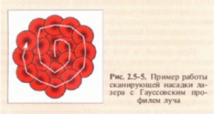
Selective photothermolysis technique
Source estet-portal.com
Share:


Add a comment