
Treatment of skin lesions in diabetes should begin with effective control of blood sugar levels. Without correction of carbohydrate metabolism, without developing an adequate regimen for taking antidiabetic drugs, all therapeutic measures related to the skin are ineffective in this group of patients.
Severe metabolic disorders underlying the pathogenesis of diabetes mellitus (DM) lead to changes in almost all organs and tissues of the body, including the skin.
Peculiarities of skin lesions in DM
The etiology of skin lesions in DM is certainly associated with a violation of carbohydrate metabolism and the accumulation of the corresponding products of disturbed metabolism, which leads to structural changes in the dermis, epidermis, follicles and sweat glands. In combination with diabetic polyneuropathy, micro- and macroangiopathies, impaired local and general immunity, this leads to the appearance of various types of rashes, age spots, ulcerations, as well as purulent-septic complications.
The skin of diabetic patients undergoes peculiar general changes. With a severe course of the disease, it becomes rough to the touch, its turgor decreases, significant peeling develops, especially of the scalp. Hair loses its shine. Calluses and cracks appear on the soles and palms. Often develops a pronounced yellowish coloration of the skin. The nails are deformed and thickened due to subungual hyperkeratosis. Diffuse hair loss can be a symptom of poorly controlled diabetes.
Often, dermatological manifestations can act as "signal signs" of diabetes: itching of the skin, dryness of the mucous membranes and skin, recurrent skin infections (candidiasis, pyoderma).
Types of diabetic dermatoses
Currently, more than 30 types of dermatoses have been described that either precede DM or develop against the background of a manifest disease. Conventionally, they can be divided into 3 groups:
1. Primary — caused by diabetic angiopathy and metabolic disorders (diabetic dermatopathy, lipoid necrobiosis, diabetic xanthomatosis, diabetic blisters, etc.).
2. Secondary — fungal and bacterial infections.
3. Dermatoses caused by drugs used in the treatment of diabetes (eczematous reactions, urticaria, toxidermia, post-injection lipodystrophy).
As a rule, diabetic skin lesions have a long and persistent course with frequent exacerbations and are difficult to treat.
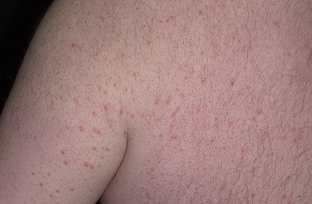
Diabetic dermatopathy. The most common lesion in diabetes is expressed in the appearance of symmetrical reddish-brown papules with a diameter of 5-12 mm on the anterior surface of the legs, which then turn into pigmented atrophic spots (more often detected in men with a long duration of diabetes). There are no subjective symptoms, the course is long, they can disappear on their own within 1–2 years. The pathogenesis is associated with diabetic microangiopathy. There is no specific treatment for dermatopathy.
Diabetic bladder. Refers to rare skin lesions in diabetes. Blisters appear suddenly, without redness, on the fingers and toes, as well as on the foot. Sizes vary from a few millimeters to several centimeters. The cystic fluid is clear, sometimes hemorrhagic and always sterile. In most cases, blisters heal without scarring after 2-4 weeks of symptomatic treatment.
Rubeosis. In childhood and adolescence, in patients with insulin-dependent diabetes mellitus, on the skin of the forehead, cheeks (less often — chin) there is hyperemia in the form of a slight blush, which is sometimes combined with thinning of the eyebrows.
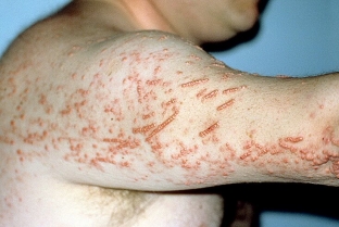
Diabetic erythema. It proceeds as ephemeral erythematous patches, which are observed mainly in men over 40 years of age with diabetes for a short time. These spots are characterized by large size, sharp borders, rounded outlines and rich pink-red color. Localized mainly on open skin — face, neck, back of the hand. Subjective sensations are either absent, or patients complain of a slight tingling sensation. The spots have a very short lifespan (2-3 days), disappear spontaneously.
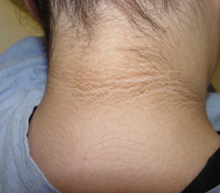
Acanthosis nigricans. Characterized by villous, hyperpigmented growths predominantly in the folds of the neck and axilla. Patients complain of "dirty skin" that cannot be washed off. On the most protruding points of the joints of the fingers, there are sometimes also small papules. The pathogenesis is based on the production of insulin-like growth factors by the liver, which bind to epidermal receptors and cause thickening of the epidermis and hyperkeratosis.
Diabetic xanthoma. It develops against the background of hyperlipidemia, and the main role is played by an increase in the content of triglycerides in the blood. Yellowish plaques are localized mainly on the flexor surfaces of the limbs, on the chest, face, neck and consist of an accumulation of triglycerides and histiocytes.
Lipoid necrobiosis in DM
A relatively rare chronic dermatosis characterized by focal disorganization and lipid degeneration of collagen.
Insulin-dependent diabetes mellitus is the most common cause of necrobiosis lipoidis and occurs in 1-4% of such patients. Skin manifestations may be the first — and for a long time the only — manifestations of diabetes. It is believed that in 18-20% of patients lipoid necrobiosis may occur 1-10 years before the development of typical symptoms of diabetes, in 25-32% of patients it develops simultaneously with this disease, but in most (55-60%) DM precedes skin lesions. There is no direct relationship between the severity of clinical manifestations of necrobiosis lipoidis and the severity of diabetes.
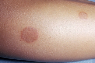
The disease can occur at any age, but more often people from 15 to 40 years old (mainly women) are affected. It occurs against the background of insulin-dependent diabetes and is characterized by large single lesions on the skin of the legs. The disease usually begins with the appearance of small bluish-pink spots or smooth flat nodules of rounded or irregular outlines, prone to peripheral growth, followed by the formation of clearly demarcated, elongated oval or polycyclic indurative-atrophic plaques. Their central part (yellowish-brown) slightly sinks, and the marginal part (bluish-red) rises somewhat. The plaques have a smooth surface, sometimes flaky along the periphery. Gradually, the central part of the plaques atrophies, telangiectasias appear on it, mild hyperpigmentation, sometimes ulceration. As a rule, there are no subjective sensations. Pain occurs with ulceration.
The appearance of the foci is so characteristic that usually no further investigation is required. In atypical forms, a differential diagnosis is made with granuloma annulare, sarcoidosis, xanthomatosis.
There is currently no effective treatment. Means that normalize lipid metabolism are used (Lipostabil, Clofibrate, Benzaflavin); improving microcirculation (Curantil, Trental, Teonikol). Such drugs as Aevit, Dipromonium, Nicotinamide, Angiotrophin are shown. Effective intralesional administration of corticosteroids, insulin, Heparin. Outwardly: applications of a 25-30% solution of Dimexide, application of Troxevasin, Heparin ointments, application of occlusive dressings with fluorine-containing corticosteroid ointments. Physiotherapy: hydrocortisone phonophoresis, Aevit, Trental electrophoresis.
Laser therapy: in case of ulceration, they sometimes resort to surgical intervention (removal of lesions with subsequent skin grafting).
Itchy dermatoses
Skin itching, neurodermatitis are often the first signs of diabetes. However, there is no direct relationship between the severity of diabetes and the intensity of itching. On the contrary: it is noted that the most severe and persistent itching is observed in latent and mild forms of diabetes. In most patients, pruritus precedes the development of not only skin lesions in DM, but also the diagnosis itself (from 2 months to 7 years). Less commonly, itching develops already against the background of established and treated diabetes.
The predominant localization is the folds of the abdomen, inguinal, intergluteal, ulnar. Lesions are often unilateral.
Fungal skin lesions in diabetes
The most common cause is candidiasis, usually caused by Candida albicans. It is more common in the elderly and in obese patients with predominant localization of foci in the genital area and large folds of the skin, interdigital folds, mucous membranes (vulvovaginitis, balanopastitis, angular cheilitis). Candidomycosis may play the role of a "signal symptom" of diabetes.
Candidiasis of any localization begins with severe and persistent itching, later objective signs of the disease join it. First, a whitish strip of macerated stratum corneum appears in the depth of the fold, and surface cracks and erosions form. Erosion surface — moist, shiny, bluish-red, bordered by a white rim. Around the main focus, "screenings" appear, represented by small superficial vesicles and pustules. Opening up, these elements turn into erosion, also prone to growth and merging. Diagnosis is confirmed by microscopic or culture examination.
For local treatment, time-tested, simple and affordable means are used — alcohol or water (the latter are better for large folds) solutions of aniline dyes: methylene blue (2 & ndash; 3%), brilliant green (1%), as well as Castellani liquid, ointments and pastes containing 10% boric acid. Of the local antimycotics, you can use almost any in the form of 1-2% creams, ointments, solutions. External agents are used until the skin lesions are completely resolved, and then for another week. Of the systemic antimycotics, fluconazole, itraconazole or ketoconazole are used. Fluconazole is prescribed at a dose of 150 mg / day once, with a torpid course, at 150 mg / day once a week for 2-3 weeks. Itraconazole is prescribed 100 mg/day for 2 weeks or 400 mg/day for 7 days. Ketoconazole is prescribed at 200 mg / day for 1-2 weeks. The expediency of prescribing systemic antimycotics is determined by the effectiveness, previous therapy, the motivation of the patient who wants to get rid of the manifestations of the disease as soon as possible, as well as the availability of drugs.
Infectious skin diseases in diabetes
Bacterial skin lesions are much more common in diabetic patients than in the general population and are difficult to treat. Diabetic foot ulcers are the most serious complication and can lead to limb amputation and even death.
Pyoderma, boils, carbuncles, phlegmon, erysipelas, paronychia and felons are most often caused by staphylococcal and streptococcal flora. The addition of infectious and inflammatory diseases of the skin, as a rule, leads to severe and prolonged decompensation of diabetes and increases the body's need for insulin. The diagnosis should be confirmed by culture and antibiotic susceptibility testing. The patient is prescribed oral dicloxacillin or erythromycin (if allergic to penicillin). Taking Dicloxacillin — the main method of treatment for outpatients, since 97% of microorganisms are sensitive to it. Lesions that have not festered can also be treated with topical heat. When fluctuating, the boil must be opened and drained. Large abscesses sometimes require incision and drainage.<
In conclusion, it should be noted that skin lesions in diabetes are today common conditions that are quite common in clinical practice. Their treatment has certain difficulties and should begin with effective control of blood sugar levels and the development of an adequate regimen for taking antidiabetic drugs. Without correction of carbohydrate metabolism in this group of patients, all therapeutic measures are ineffective.According to http://www.lvrach.ru/
Share:


Add a comment