


Actinomycetes – amazing microorganisms that are an intermediate form between fungi and bacteria.
They have a cell wall and structure similar to Gram-positive bacteria and characteristic branching filaments – mycelium, like mushrooms. Actinomycetes are common saprophytes in humans and animals, vegetating in the mouth and intestines.
Under certain conditions, microorganisms cause the disease actinomycosis. On estet-portal.com read about the main causes, types and symptoms of actinomycosis, as well as about the diagnosis and treatment of the disease.
Main causes, routes of infection and clinical forms of actinomycosis
Actinomycosis – chronic specific inflammatory disease caused by radiant fungi – actinomycetes.
Together with erythrasma, actinomycosis belongs to the group of pseudomycoses. A person becomes infected by endogenous (autoinvasion) or exogenous (consumption of contaminated cereals and water) by.
Microtraumas, cracks, carious teeth in the oral cavity contribute to the deep penetration of actinomycetes into the body.
Pyogenic microflora also creates the conditions – enzymatic background – for the development of actinomycosis.
The following forms of actinomycosis are distinguished by clinical manifestations:
• knotty;
• fungal-pustular;
• subcutaneous facial.
According to the localization of the disease:
• cervicofacial;
• bronchopulmonary;
• intestinal;
• other forms (any organs are affected).
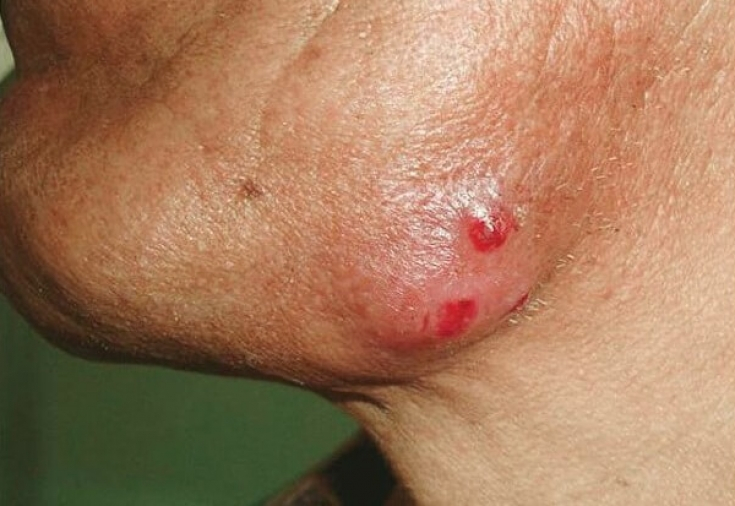
The most common is the cutaneous-facial form of actinomycosis, which occurs in 60% of cases.
Follow us on
Instagram
The infiltrate acquires a pink-cyanotic color and gradually softens due to necrosis in its center.
Soon an abscess develops, which breaks out in several fistulas with pus.
In severe stages of actinomycosis, masticatory muscle contracture develops, inflammation of the subcutaneous fat and osteomyelitis.
 The intestinal form of actinomycosis may resemble acute appendicitis or intestinal obstruction, with palpation of the abdomen, tumor-like masses can be felt.
The inflammatory process can spread to the mediastinum and other organs with pronounced signs of intoxication of the body.
Erythrasma: a disease that benefits from the sun
Methods of diagnosis of actinomycetes and approaches to the treatment of actinomycosis
Treatment of actinomycosis is complex: conservative-surgical. In the presence of significant purulent processes on the skin, the abscess is opened and drained.
Systemically used benzylpenicillin in high doses (20 million OD after 6 hours).
In the presence of resistance to penicillins, tetracycline antibiotics, cephalosporins or aminoglycosides are used.
Therapy with actinolysate (immunomodulator) is carried out intradermally or intramuscularly.
The course of treatment is 3 months twice a week at a dose of: 0.5-3 ml with a gradual increase in dose.
Iodine preparations, in particular iodonate, are applied locally.
Actinomycosis, provided timely diagnosis and adequate treatment, relatively rarely leads to fatal consequences.Thank you for staying with estet-portal.com. Read other interesting articles in the "Dermatology" section. You may also be interested in:
Main aspects and methods of treatment of actinomycosis