

Wounds and related injuries remain the leading cause of death and disability. The healing process includes 3 phases: inflammation, proliferation, tissue formation and remodeling.
The proliferative phase includes re-epithelialization and formation of granulation tissue, which includes fibroplasia and angiogenesis.
Keratinocytes migrate 12-24 hours after injury. Migration and proliferation of these cells are key events for re-epithelialization and wound closure. During the formation of granulation tissue, fibroblasts migrate, proliferate, and synthesize large amounts of collagen and other extracellular matrix to fill in the skin defect in a process known as fibroplasia. Angiogenesis depends on the migration and proliferation of endothelial cells from pre-existing blood vessels in the wound.
Aloe Vera Leaf contains chemical compounds (e.g. acetylated mannans, polymannans, anthraquinone C-glycosides, anthrones, anthraquinones and lectins) and is traditionally included in many cosmetic and alternative medicines for anti-aging, wound healing and other dermatological conditions.
In the article estet-portal.com you can get information regarding the physiological function of aloe vera in wound repair.
Materials and methods for studying the effectiveness of Aloe vera in wound healing
The experimental liquid contained 90% Aloe Vera (internal gel), deionized water, and preservatives consisting of 0.1% sodium benzoate, 0.1% potassium sorbate, and 0.14% phosphoric acid. Various concentrations of Aloe Vera solutions were prepared at 1%, 2% and 3%.
Follow us on Instagram!
For fibroblasts, the fluid was diluted in Modified Dulbecco's Medium with 2% fetal bovine serum. For keratinocytes, the liquid was diluted in keratinocyte growth medium. Also, since Aloe Vera liquid contained preservatives (mixed with Aloe Vera product), the same culture media with preservatives but without Aloe Vera were included as controls.
Human primary epidermal keratinocytes and dermal fibroblasts have been isolated from human neonatal foreskin.
Cells were divided into 4 groups, scratched, and then treated with 1%, 2%, and 3% Aloe Vera every 24 hours from day 0 to day 5 of the experiment. Proliferation results were compared with control (Ctr).
Watch the most interesting videos on our
The effect of Aloe vera on fibroblast proliferation was evaluated in a 5-day course. As shown in Figure 1, 2% concentrations of Aloe Vera solution showed a strong stimulatory effect on fibroblast proliferation compared to 2% control medium (P < 0.05 on day 1, P < 0.01 on days 2 and 3 and P <0.001 on day 5).
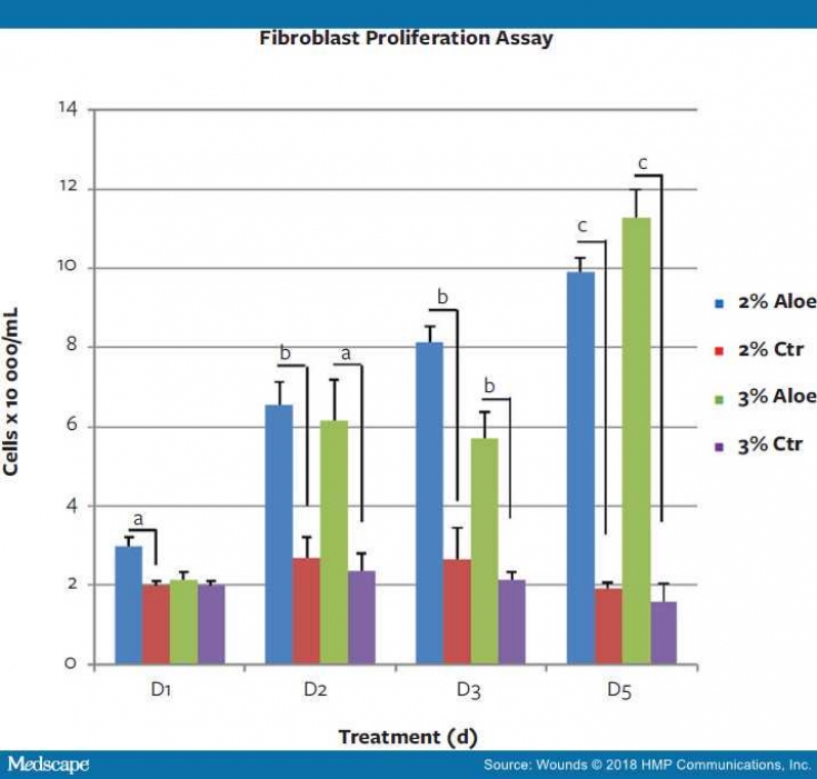
Graphic analysis of the average number of fibroblast cells. Cells treated with aloe vera (2% aloe and 3% aloe) were compared with the corresponding control media (2% Ctr and 3% Ctr).
Aloe vera stimulated fibroblast proliferation.
Effect of Aloe Vera on Fibroblast Migration: Fibroblasts treated with 3% Vera experienced accelerated gap filling (29%) compared to 3% Ctr medium (17%) within 24 hours (P < 0.05).
Aloe Vera stimulated fibroblast migration.
What is the secret to fast wound healing How Aloe Vera stimulates keratinocytes
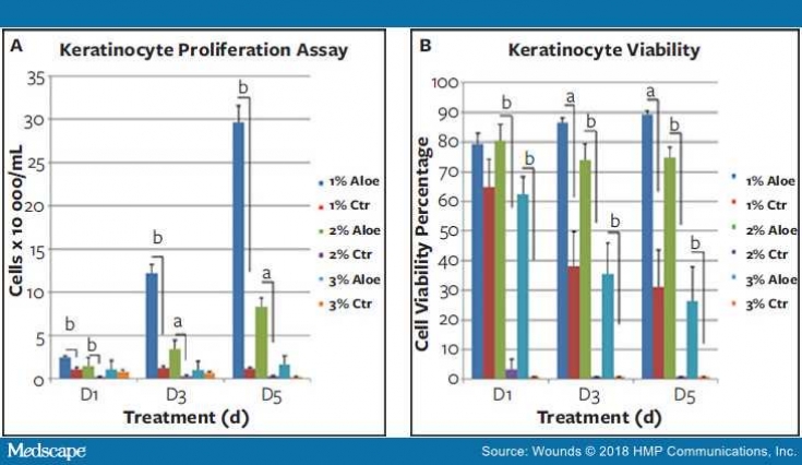
Throughout the study, both 1% and 2% concentrations of Aloe vera showed a very strong stimulatory effect on keratinocyte proliferation compared to Ctrs with P<0.01 or P<0.001 over all 3 days of the study (A) . Aloe vera 1% had better effects than Aloe vera 2% throughout the study, and Aloe vera 3% had no significant effects. Graphical analysis of mean viable cells (B) shows that Vera increases the viability of keratinocytes.
Aloe Vera stimulates the proliferation of keratinocytes and increases the viability of keratinocytes.
Keratinocyte migration assay results showed that Aloe vera at 1% had a better effect on cell migration as measured by PGF 24 hours after scratching compared to 1% Ctr medium (P <0.05).
Aloe Vera stimulated the migration of keratinocytes.
Aloe Vera: scientifically proven efficacy
While this study demonstrated the potential mechanisms of Aloe vera to promote wound healing in cell proliferation and migration, in vitro wound healing assays cannot mimic the complexity of the conditions that occur during the in vivo wound healing process. Therefore, data obtained from in vitro assays should not be considered conclusive and should be confirmed by in vivo models.
Thank you for staying with estet-portal.com. Read other interesting articles in the "Dermatology" section. You may be interested in
The role of cytokines in the treatment of non-healing wounds
Adapted from Medscape
Share:


Add a comment