

Transepidermal water loss (TEWL) is the most widely used objective measurement to assess skin barrier function in healthy individuals as well as patients with skin conditions such as atopic dermatitis (AD).
A stronger skin barrier is characterized by larger superficial corneocytes, an increased number of corneocyte layers, and/or improved intercellular lamellar lipid matrices.
The molecular organization of the extracellular lipid matrix of PC in a highly ordered lamellar bilayer structure is an important determinant of TEWL.
In the article estet-portal.com you can get acquainted in detail with the reason for the increase in TPPV in atopic dermatitis and the possibilities of using this indicator for the early diagnosis of impaired skin barrier function.
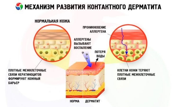
Impaired skin barrier in atopic dermatitis
Dysfunction of the skin barrier leads to an increase in TEWL. Skin diseases in which the skin barrier is compromised, such as atopic dermatitis, contact dermatitis, psoriasis, and ichthyosis, are associated with increased TEWL. In addition to in vivo measurement of water barrier function, TEWL measurement correlates with transdermal absorption of pathological factors (allergens, toxins).
TEWL measurements can be thought of as an indirect measure of skin permeability, reflecting the function of the protective barrier state.
TEWL is used as a research tool to objectively assess skin barrier function and it can be reliably correlated with atopic dermatitis severity and response to treatment, leading to its inclusion in some AD severity assessments.
Barrier dysfunction and increased TEWL are the main pathological features of atopic dermatitis.
Watch the most interesting videos on our channel in Youtube
Measurement of transepidermal water loss for the diagnosis of atopic dermatitis
Filaggrin is a key component of the epidermal skin barrier, and up to 50% of patients with moderate to severe AD carry one of the mutations associated with a defect in the functional filaggrin gene (FLG).
Studies have shown that at birth there is no difference in TEWL between FLG mutation and normal FLG groups. However, at 2 months, 3 months, and 6 months, those with the FLG mutation have a significantly higher TEWL than people without the filaggrin gene mutation.
TEWL has been found to be elevated in infants with FLG-null mutations without clinical AD, indicating that skin barrier disruption may precede clinical manifestation of AD.
It has been shown that TEWL measured in the first days of life can predict the development of AD in infancy, regardless of FLG status.
These data suggest that TEWT has the potential to be used to identify neonates at increased risk of developing AD and to help guide prevention strategies, such as with the use of a conventional emollient.
Thank you for staying with estet-portal.com. Read other interesting articles in the "Cosmetology" section. You may be interested in New options for the treatment of atopic dermatitis in children and adults
Share:


Add a comment