
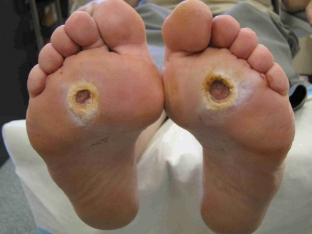
The presence of diabetes sooner or later affects the functioning of many body systems, as well as the appearance of the patient. In 10% of patients with diabetes mellitus, there are disease-specific changes in foot tissues caused by metabolic disorders in the body. Such patients constantly feel pain in the legs, their distal limbs are deformed, soft tissue necrosis and ulcerative defects, skin cracks and hyperkeratosis appear. These changes are called "diabetic foot". What is the mechanism of development of diabetic foot? What does a diabetic foot look like in a photo?
When does diabetic foot development begin? Photo of a diabetic foot
Diabetic foot usually develops 15-20 years after the first manifestations of diabetes mellitus (DM) in a patient. At the present time, the treatment of diabetic foot is an urgent issue, since in most cases such a complication of diabetes is detected in the later stages, which dictates the need for amputation of the limb. This leads to disability of patients, a decrease in the quality of life and an increase in mortality. Therefore, the treatment of diabetes mellitus should prevent the development of diabetic foot.
To do this, it is important to understand the mechanism of development of pathological changes in the foot. The main pathogenetic links in the development of diabetic foot are neuropathy, angiopathy and infection.
Mechanism of development of diabetic foot in patients with DM
Long-term hyperglycemia leads to pathological changes in blood vessels and peripheral nerves. Angiopathy is accompanied by a decrease in the elasticity and strength of blood vessels, an increase in blood viscosity. This, in turn, leads to disruption of innervation and trophic processes in the tissues, so they lose sensitivity.
Intensive glycosylation of proteins provokes a decrease in joint mobility, which leads to deformation of the bones of the limb. The biomechanical load on the foot is disturbed (Charcot foot). The slightest injury to the foot against the background of reduced sensitivity, impaired blood circulation and a decrease in the protective function of the tissues of the foot leads to the formation of ulcers that do not heal for a very long time.
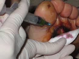
Tissue defects in diabetic foot (photo) often lead to & nbsp; infection of the body with anaerobic flora, streptococci, staphylococci and colibacilli.
 Bacteria secrete bacterial hyaluronidase, which, loosening the surrounding tissues, creates conditions for the spread of the infectious process. Thus, subcutaneous fat, muscle tissue and bone-ligamentous apparatus are exposed to infection and necrotic changes. In this case, there is a high risk of developing phlegmon, gangrene or abscess of the limb. The risk of developing a diabetic foot exists in every diabetic patient, but a risk group is distinguished.
Factors that form the risk group for developing diabetic foot in diabetes mellitus:
- vascular atherosclerosis;
- hyperlipidemia;
- ischemic heart disease;
- arterial hypotension;
- peripheral polyneuropathy;
- arterial hypertension;
- bad habits (drinking alcohol, smoking). ***
What can provoke the development of a diabetic foot (photo)?
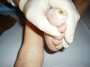
Local tissue changes in the leg also increase the likelihood of developing a diabetic foot (photo): these are ingrown nails, skin and nail mycoses, cracks and calluses, as well as poor foot hygiene.
The reason for the development of diabetic foot may be improperly selected shoes. With tight or narrow shoes, the patient does not feel pain and discomfort due to a decrease in sensitivity, while the foot is injured, and conditions are created for the development of a diabetic foot.
 The diabetic foot is a serious pathology, which, first of all, must be prevented by adequate treatment of diabetes mellitus and related pathologies. It is also necessary to explain to the patient the importance of regular personal hygiene and foot care. When the first signs of the process are detected, it is necessary to start treating the diabetic foot (photo).
Share:


Add a comment