


Tilapia skin (a species of fish) has a non-infectious microbiota, abundant type I collagen and a morphological structure similar to human skin, therefore it has been proposed as a potential xenograft for the treatment of burns ran.
A 23-year-old patient arrived at a burns treatment center in Brazil after suffering a thermal injury caused by contact with fire from a gunpowder explosion.
Both of his hands were injured from deep burns, with the left being particularly affected.
On estet-portal.com read how a patient was treated with tilapia skin and what results this therapy allowed to achieve.
- Tilapia skin has been used to treat superficial and deep burns
- Clinical case: application of tilapia skin on a real patient
- Tilapi burn healing processand
Tilapia skin has been used to treat superficial and deep burns
According to World Health Organization, burns cause 180,000 deaths each year, most of which occur in low- and middle-income countries, of which Brazil is a group.
Furthermore, non-fatal burns cause prolonged hospitalization, disfigurement and disability, combined with stigmatization.
Tilapia skin Nile Tilapia Fish Skin (NTFS) has been proposed as a biological material for treatment burns.
Colony forming units found in NTFS samples prior to the chemical sterilization process indicate the presence of normal non-infectious microbiota.
Tilapia skin was found to be rich in type I collagen, morphologically similar to human skin, and also highly resistant to tensile strength.
Read the most interesting articlesand in Telegram!
Clinical case: application of tilapia skin on a real patient
A 23-year-old patient suffered a thermal injury caused by contact with fire from a gunpowder explosion.
Burns accounted for more than 16% of total body surface area.
After admission to the hospital, he was resuscitated and remained hemodynamically stable.
Tilapia skin was applied to lesions resulting in complete re-epithelialization within 12 (superficial burns) and 17 (deep burns) days of treatment.
No side effects observed.
Treatment of first degree burn after chemical peel: case report
Tilapia skin promises to be an innovative, easy-to-use and widely available product for burn therapy.
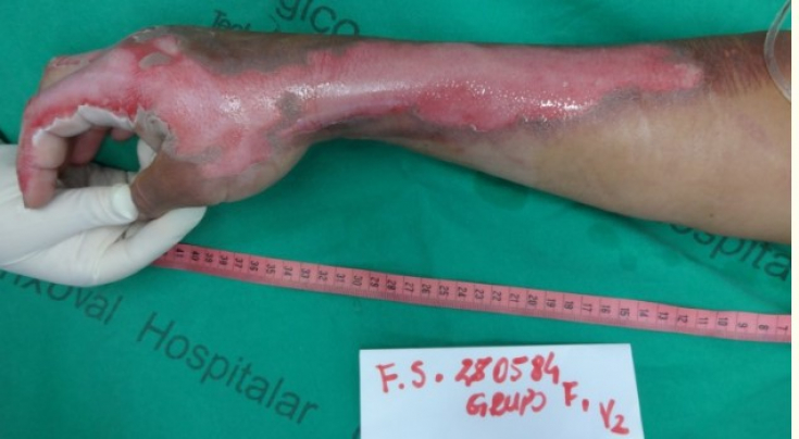
Figure 1: Superficial burns on the right hand
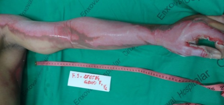
Figure 2: Deep burns on the left hands
The process of healing burns under the skin of tilapia
Tilapia skin has undergone a rigorous process of chemical sterilization, glycerolization and irradiation followed by microbiological testing for bacteria and fungi before being stored in refrigerated sterile packaging.
Prior to use, the patient's skin was flushed with sterile 0.9% saline for 5 minutes, and this process was repeated three times in a row.
Tilapia skin did not change its microscopic and tensometric structure after chemical sterilization and irradiation, and also restored its natural texture after the rehydration process.
The patient was subjected to anesthesia and analgesia.
Surgical treatment of burns: necrectomy operation
After cleaning the lesion and removal of necrotic and fibrinous tissues (Fig.3) the biomaterial was applied to the upper limbs of the patient (Fig.4).
When applying the graft, at least 1 cm of healthy skin at the wound margins must be covered with a distance of at least 1 cm between the NTFS pieces to ensure that possible movements in the first days of treatment do not lead to the opening of any area of the burn.
Finally, the wounds were covered with a gauze bandage.
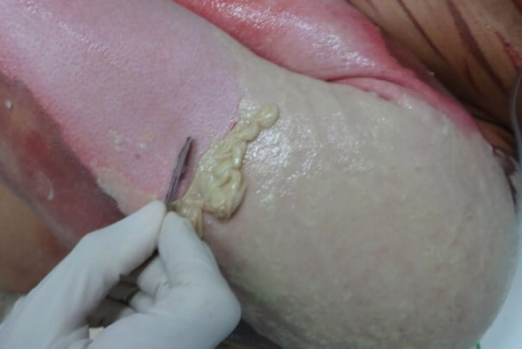
Figure 3: The process of removing necrotic and fibrinous tissue from a lesion
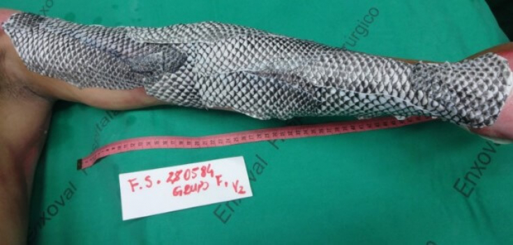
Figure 4: Appearance of the left upper limb after applying NTFS.
Daily, the patient's blood was taken for analysis, and no significant changes were found.
Also, the patient's vital signs and clinical condition remained stable.
Even deep burns are not terrible with competent surgical tactics
The biomaterial showed good adhesion to the wound bed without the need to change the dressing (Fig.5).
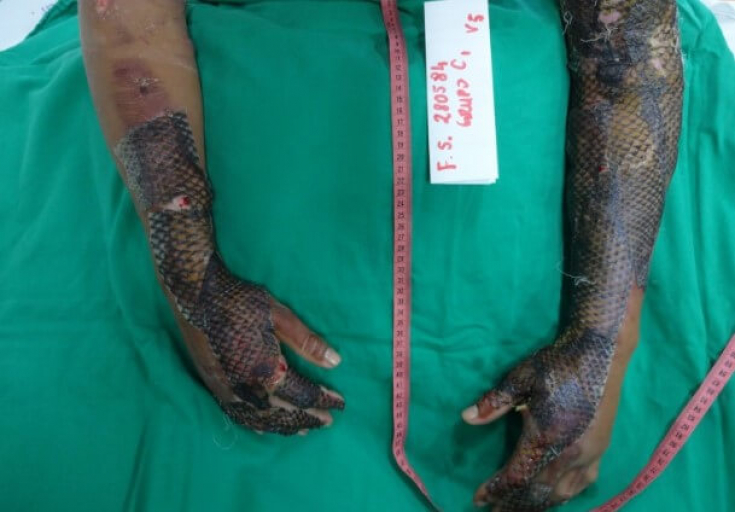
Figure 5: Skin appearance on the sixth day of treatment. Good adhesion of NTFS to the wound bed.
On the 12th day of treatment the skin of the tilapia appeared dry and hardened and began to come off the patient's right upper limb.
So the researchers decided to remove NTFS from the area.
Hand was treated with water, causing the telapia skin to separate, revealing healed skin (Fig.6).
, the skin of the tilapia was also removed from the left arm, which initially had deep burns (Figure 7).
No side effects reported.
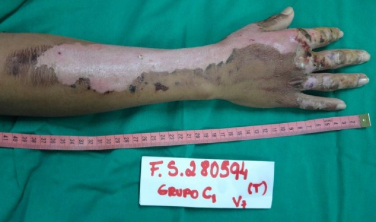
Figure 6: Right arm after NTFS removal (12 days from start of treatment)
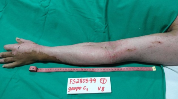
Figure 7: Left arm after NTFS removal (17 days from start of treatment) The use of NTFS as a xenograft for the treatment of experimental burns in rats resulted in an improvement in the healing process and the absence of corresponding changes in hematological and biochemical parameters.
A recent study has shown thattilapia collagen significantly induces epidermal growth factor
and fibroblast growth factor expression, which may promote the proliferation and differentiation of fibroblasts and keratinocytes.
Surgical treatment of burns: graft harvesting The periods of 12 and 17 days required, respectively, for
complete re-epithelialization of the woundsof this patient indicate the efficacy of tilapia skin as a xenograft for the treatment of human burns. In order to introduce this method into general practice,
more extensive studiesare naturally required, on which Brazilian scientists have already begun work.