Laser is quite effective in treating telangiectasias with little or no risk of subsequent scarring or other permanent skin changes.
Telangiectasia – this is an expansion of superficial vessels (venules, capillaries, arterioles) with a size of 0.1-1.0 mm in diameter. Clinically, telangiectasia is divided into four groups:
- simple or linear;
- branched;
- arachnid;
- papular.
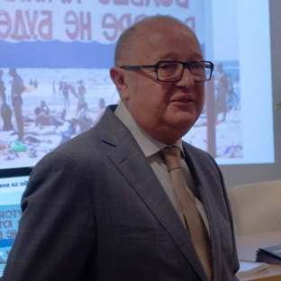
Vladimir Tsepkolenko told Estet-portal about the methods of treatment telangiectasias.
 Vladimir Alexandrovich Tsepkolenko
MD, Professor, Honored Doctor of Ukraine,
President of the Ukrainian Society of Aesthetic
Medicine, General Director of the Ukrainian
Institute of Plastic Surgery
and aesthetic medicine "Virtus"
Red linear and branched telangiectasias are often found on the face, especially in the nose, central part of the cheek, chin, and legs. Blue linear and branched telangiectasias most often occur on the legs, manifestation in the face is not excluded. Papular (nodular) telangiectasia is often part of genetic syndromes such as Osler's disease – Weber – Rendu, and is also observed in vascular collagenosis.
Linear facial telangiectasia
Traditional methods of treatment of large vessels of the alar nose often lead to noticeable scarring. The dye laser is quite effective in treating this type of formation with little or no risk of subsequent scarring or other permanent skin changes.
When removing superficial vessels up to 0.3 mm in diameter, yellow-green spectrum radiation (KTP 532 nm, PDL 575-600 nm) is effective, however, deeper vessels (or vessels of large diameter) are not amenable to the action of these lasers due to insufficient depth penetration of their radiation. Therefore, when processing deeply located vessels with a diameter of up to 1.5 mm, lasers with a long wavelength (diode, 800-1000 nm, Nd:YAG, 1064 nm) are used. The end point of the procedure is the disappearance of the vessel, and not discoloration, blistering or charring of the upper layer of the skin.
The main side effect – hyperpigmentation, but it is unlikely to occur.
Arachnoid hemangioma (spider vein, spider nevus, stellate hemangioma)
This is a benign vascular tumor that is a variant of capillary hemangioma. It is localized more often on the face, forearms and hands. Its appearance may be associated with an excess of estrogens (pregnancy, taking oral contraceptives), with damage to hepatocytes (hepatitis, cirrhosis of the liver). Most often formed at the age of 7-10 years. May occur in healthy people (up to 15%). In the center of the neoplasm there is a small (several millimeters) red papule (feeding arteriole), from which dilated convoluted capillaries extend. The general sizes of a hemangioma – 1-1.5 cm.
Treatment. High efficiency is shown by the dye laser and the KTP laser. In 70% of cases, formations are completely removed in one session, almost all remaining – during a repeat procedure. The standard approach assumes a radiation energy density of 6 to 7.5 J/cm2. One or two impulses are directed to the central part of the "spider", and the rest are superimposed with a 10% overlap on the "paws". damage if its diameter exceeds a few millimeters. Immediately after treatment, the formation becomes purple-red, but within 1-2 weeks the redness disappears on its own. Local anesthesia is usually not used. The likelihood of permanent pigment changes or scarring is minimal.
Poikiloderma (poikiloderma Civatta) and its treatment
This variant of telangiectasia, involving the neck and upper chest, is the result of long-term ultraviolet exposure. Poikiloderma combines telangiectasia, pigmentation disorders and atrophic skin changes. These changes are best treated if the vascular and pigment components are simultaneously affected. When treating the vascular component, quite good results are possible, but often a large number of procedures are required. Successful application of the KTP laser (532 nm) has been reported. Argon laser often leaves areas of hypopigmentation with some potential for scarring.
Rosacea: methods of exposure and effectiveness of treatment
At its core, rosacea – these are cutaneous and vascular disorders including telangiectasia, papules, pustules and rhinophyma; most often develop at the age of 30-60 years, although cases of diagnosis of this disease in children are known.
Comprehensive treatment for rosacea includes:
- drugs to eliminate digestive dysfunction, hormonal correction, possibly – antibacterial drugs;
- external therapy;
- proper nutrition (often, fractionally, exclude fried, smoked, spicy, very hot dishes, alcohol);
- avoid thermal procedures (bath, sauna, solarium, paraffin therapy, vaporization);
- correct cleansing of the skin with cosmeceuticals;
- ultrasound skin therapy with drugs;
- laser therapy.
Like all other types of telangiectasia, rosacea can be treated with vascular lasers (KTP and PDL), but their effectiveness is not high enough. We have accumulated a lot of experience using QuadroStar + Nd:YVO4 (wavelength 532 nm, spot size 1.5 mm, power 5 W), as a rule, 3-5 treatments are enough, depending on the stage of the process.


Add a comment