

Hyaluronic acid fillers are most often used for contouring due to their high safety profile. Despite this, no practitioner is immune from serious complications of injection correction. The article estet-portal.com presents a case of upper lip tissue necrosis after fillera HA injection from the practice of Dr. Peter Hirsch (Peter Hirsch), Manfred Infanger (Manfred Infanger) and Armin Kraus ( Armin Kraus). The authors talk about the causes of necrosis after the introduction of fillers, and also provide recommendations for management and prevention of this complication.
- Case report: necrosis of upper lip tissues after HA injections
- Tissue necrosis after fillers injection: causes, therapy and prevention of complications
Tissue necrosis of the upper lip after HA injections: a case report
A 19-year-old female patient presented to the authors' clinic one week after a filler injection into her upper lip. What kind of product was administered to the patient and in what quantities is unknown. According to the patient, the procedure was performed after applying an anesthetic cream. Despite this, during the injection, the girl felt a strong burning pain that radiated from the left side of the upper lip to the nose. Other than cooling compresses, no specific post-contouring therapy was prescribed.
Follow us on Instagram!
During the examination, a dry necrotic area measuring 8 x 10 mm was found, as well as swelling of the left part of the upper lip. From the upper lip through the filtrum and columella to the tip of the nose, a line of a blue tint was visualized.
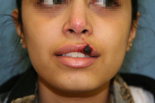
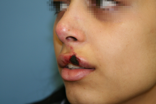
Photo 1: Partial necrosis of the left side of the upper lip and impaired perfusion of the skin of the filtrum, columella and tip of the nose after HA injection
Therapy
Since a lot of time has passed since the procedure, the introduction of hyaluronidase was inappropriate. The authors decided to remove as much of the filler as possible located under the area of tissue necrosis.
The patient underwent a blockade of the infraorbital nerve, a small stabbing incision was made on the mucosal side, and the filler was removed.
You may also be interested in: A case of occlusion of the posterior ciliary artery after injection of hyaluronic acid into the forehead area
Dexpanthenol ointment was prescribed as a topical therapy, as well as regular application of dry dressings for 2 weeks.
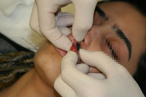
Photo 3: Filler removal after infraorbital nerve block
After 3 months the wound healed, and a small scar formed in its place. There was also a significant loss of volume in the left side of the upper lip. The color of the skin of the affected areas was almost restored to normal, however, thickening was felt in the tissues, which indicated subdermal scarring. The asymmetry was enhanced by the presence of a filler on the right side of the upper lip. Causes, treatment and prevention of complications of tissue necrosis after fillers
Tissue necrosis after filler injection is more often the cause of intravascular injection of material and occlusion of supply vessels as a result of embolization.
Most often, the complication develops after intravascular injection of the filler into:
nasolabial fold (facial artery);
- intereyebrow (trochlear artery);
- nose (angular artery). The retrograde movement of the filler through the collateral vessels into the terminal branches is a consequence of the injection of large volumes of the drug into the artery under high pressure.
The pattern of tissue injury may depend to some extent on the vascular anatomy (affected vessel and degree of collateralization) and
physical properties of the injected filler
(viscosity/particle size). Since the facial vasculature is well developed and skin necrosis develops in areas with severe collateralization, it is assumed that filler occlusion occurs at the capillary level, where collateralization is not able to compensate for impaired perfusion. An additional factor may be the hygroscopicity of the material, which increases in volume in the capillary bed, increasing endothelial damage and occlusion. Smaller filler particle size may also increase the risk of capillary occlusion to some extent in case of intravascular injection of the product.
Read also:
Principles for the safe administration of fillers in hazardous areas In the present case, the healing of the lesions resulted, as the authors suggest, in subdermal scarring with induration and moderate volume loss. It follows from this that tissue necrosis was the result of
occlusion at the level of capillaries
, even in areas where the integrity of the skin is not broken.
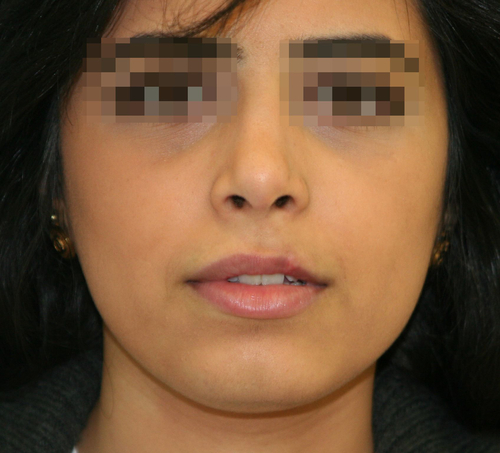
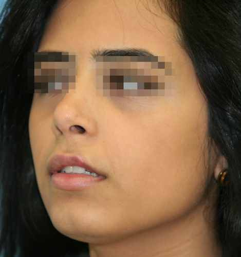
Photo 4: 3 months after therapy
Recommended therapy in case of impending necrosis after arterial filler injection:
interstitial hyaluronidase injection and massage;
- application of nitroglycerin paste;
- local heat to improve perfusion;
- hyperbaric oxygen therapy. In general, the physician's ability to influence the outcome of an intra-arterial filler injection is very limited. Therefore, the prevention of complications comes to the fore.
How to avoid getting a filler into an artery:
know anatomy well and work with extreme caution in hazardous areas;
- inject small volumes of the drug after the aspiration test;
- Use smaller syringes and needles for better injection control and less injection pressure;
- use blunt cannulas;
- inject the product anterograde and retrograde, constantly moving the cannula;
- pressure a blood vessel through which the filler can move retrogradely;
- Use epinephrine to stimulate vasoconstriction and reduce the risk of intravascular injection.
Ischemic skin necrosis after HA filler: diagnosis and management of complications The practitioner should remember that the division of the face into dangerous and safe zones is rather arbitrary – tissue necrosis can develop after the introduction of a hyaluronic acid filler at any point. Therefore, the authors strongly recommend that all safety rules be observed when performing contouring procedures, regardless of the product used.
Adapted from
Journal of Cosmetic Dermatology
. More interesting videos on our
YouTube-channel!
Share:


Add a comment