
Osteoporosis of the bone is a fairly common skeletal disease that has a systemic nature. It is characterized by a decrease in bone density, a violation of its microarchitecture, which further increases the risk of fractures. In Europe, Japan and the USA, approximately 75 million people suffer from this pathology.
The annual incidence of osteoporotic bone fractures is 9 million, of which 1.6 million are femoral neck fractures. Such osteoporotic fractures significantly affect morbidity and mortality. As a result of a fracture of the femur, mortality increases by 12-15%.
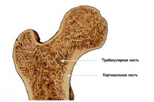
Main indicators of bone tissue in the detection of bone osteoporosis
The most difficult detection of bone osteoporosis in the early stages, since in most cases the first sign is a low-energy fracture. In this regard, in recent years, there are more and more new methods for diagnosing osteoporosis of the bone, which help to identify risk groups and early loss of bone density.
There are several factors that affect the condition of the bone tissue. These are the degree of mineralization, macrogeometry, mineral density of tissue and its metabolism, the presence of microfractures and the microarchitecture of trabecular bone tissue. The most significant factor in the development of osteoporosis of the bone is the mineral density of the tissue. The gold standard for detecting bone mineral density is two-photon X-ray absorptiometry (DXA).
The use of this method for diagnosing osteoporosis of the bone has some limitations:
- The presence of a significant "overlap zone" in people who develop fractures and in those who do not. This restriction is one of the main ones.
- Disproportionate assessment of the cortical ball of the bone depending on the area examined using DXA, and differences in the exchange in the bone tissue of the studied areas.
- During treatment, the likely changes in the indicator can be assessed after a long time, which is calculated in years. In contrast to the cortical tissue, the exchange in the trabecular bone tissue occurs several times faster. Therefore, it is the assessment of the microarchitecture of the trabecular bone that increases the sensitivity and accuracy of assessing the quality of the entire bone tissue, as well as the risk of fracture in clinical practice.
Identification of bone osteoporosis by assessing the structure of trabecular tissue
 The structure and condition of trabecular bone can be assessed using magnetic resonance imaging or computed tomography, but both techniques are expensive to use and not always available in clinical practice. Also, the assessment of the microstructure of the bone tissue can be carried out by histomorphometric analysis during a biopsy of the bone tissue of the iliac crest.
Although this diagnostic method is highly informative, manipulation is an invasive and traumatic procedure. Moreover, it is debatable how much this site will reflect the presence of osteoporosis of the bone and the risk of fracture. To date, new non-invasive imaging technologies are being developed in diagnosing disorders of the structural and functional state of bone tissue and identifying the risk of fracture.
Assessment of the quality of trabecular bone tissue in the diagnosis of bone osteoporosis
A French company has patented a new method, TBS Insight, for assessing trabecular bone quality and detecting osteoporosis. When evaluating the indicator, no direct physical measurement of the bone microarchitecture is carried out; the indicator is calculated using the 3D projection – structures on a 2D plane.
The analysis is based on the grayscale variation and pixel density amplitude of the x-ray image. The method makes it possible to analyze the trabecular structure according to different statistical properties of pixels. As a result, a score is calculated that strongly correlates with the 3D parameters of the designed trabecular bone. This method of diagnosing osteoporosis of the bone gives a global assessment of the quality of bone tissue. The importance of the technique lies in its discriminatory ability, sensitivity of indicators to changes in the disease or treatment, as well as the assessment of the risk of osteoporotic fractures.
Thus, modern medicine provides an opportunity to detect violations in the structure of bone tissue before fractures. This will significantly reduce the morbidity and mortality of the population from fractures of large bones, which is especially important in old age.
Share:


Add a comment