
Electrosurgery – a method based on the application of high-frequency electric current to biological tissues for cutting, coagulation, dissection and fulguration. The main advantage of this method is the ability to perform an accurate cut of biological tissues with minimal blood loss. Electrosurgical devices are often used during a wide variety of surgical interventions, both in hospitals and in outpatient practice.
When performing surgical interventions using electrosurgical devices, biological tissues are heated, these devices can be used to cauterize tissues. The term "electrosurgery" used to refer to a technique other than electrocautery that directly heats the tip of the device with a direct electric current.
The mechanism of interaction of a radio wave with a biological tissue is fundamentally different and lies in the fact that a high-energy radio wave, coming from a generator through an active electrode, is directed to a passive electrode (called an "antenna"), meets the resistance of the cells and instantly heats them up. As a result, the intracellular fluid boils and breaks the cell membrane. When boiling, small vapor bubbles are formed, which push the tissues apart in front of the radio wave. Let us note the characteristic feature of the radio wave, which already when approaching the surface of the tissue begins its impact.
Of great importance for a correct understanding of the mechanism of action of a radio wave instrument is the fact that during the operation the active electrode remains cold, which distinguishes it favorably from a laser beam and an electrosurgical scalpel, since it does not cause a noticeable burn of the surrounding tissues and promotes rapid wound healing with less scarring.
Modern radio wave surgical devices have the ability to generate 4 types of waves:
- "Cut" (cut) (3.8 & ndash; 4.0 MHz). In this mode, the device generates a fully straightened and filtered radio waveform, which is a continuous stream of high-frequency vibrations, producing the thinnest, perfectly even cut. Such a wave provides the least transverse heating and tissue destruction. 90% of radio wave energy is spent on the incision, 10% – for coagulation. In "Cut" mode clean, microscopically even incisions with slight coagulation are performed: cosmetic skin incisions, biopsy, opening of abscesses, removal of keratomas, formation of skin flaps, elimination of cosmetic defects and other manipulations on the skin and soft tissues that do not require enhanced hemostasis.
- "Cut/Coag" (cut/coagulation) (3.8-4.0 MHz). The fully extended waveform is a weakly pulsating radio wave producing an incision with slight superficial coagulation (no charring) – the so-called "coagulation film" on tissue cuts. Such coagulation effectively stops bleeding and seals the nerve fibers, which allows you to perform "dry incisions" and reduces pain in the postoperative period. 50% of radio wave energy is spent on the incision, 50% – for coagulation. In this mode, tissue dissection occurs simultaneously with surface coagulation. In this mode, most surgical procedures are performed on all subcutaneous tissues, mucous membranes and internal organs, neoplasms are removed: "Coag/Hemo" (coagulation / hemostasis) (3.8 - 4.0 MHz). The partially rectified waveform is a pulsating stream of high frequency vibrations. 90% of radio wave energy is spent on coagulation, 10% – for the cut. Radio wave coagulation differs from electrocoagulation in the absence of tissue charring and does not form a burn eschar.
- The spark gap wave, which is referred to as "fulguration" (from Latin fulgur – "lightning") and is used in various electrosurgical devices to stop bleeding.
Speaking about the use of radio wave devices in surgery, it should be noted that the doctor can easily learn how to work with them, ease of manipulation, reduced intervention time, no need for special operating room equipment, high safety of their use for both the patient and medical personnel.
In the clinic of diseases of the ear, nose and throat of the First Moscow State Medical University named after I.M. Sechenov, over the past few years, the Surgitron apparatus has been used the Ellman company. Extensive experience has been accumulated in the surgical treatment of a number of diseases of the ENT organs using the radio wave method. The clinic actively uses the radio wave in the surgical treatment of patients with various chronic diseases of the nasal cavity (bipolar cautery of the inferior turbinates); submucosal uvulopalatoplasty for the treatment of snoring and sleep apnea; microsurgical operations on the vocal folds; lacunotomy in the treatment of chronic tonsillitis; functional endoscopic rhinosinus surgery and in many other cases when it is required to perform an accurate and bloodless incision of biological tissues.
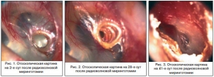
A number of experimental and clinical studies have been carried out, proving the high efficiency of using radio waves during myringotomy. In particular, in the treatment of patients with exudative otitis media, it is necessary to evacuate the pathological contents and restore the normal function of the auditory tube for good aeration of the middle ear cavities. In the treatment of this pathology, shunting of the tympanic cavity is widely used in the world. The specialized literature describes numerous complications from the use of this type of treatment, mainly associated with the long-term presence of a foreign body (shunt) in the tympanic membrane and tympanic cavity. Radio wave myringotomy is simple to perform and eliminates the need for a tympanostomy tube. Closing the tympanostomy in a short time allows you to eliminate the causes of dysfunction of the auditory tube. In our clinic, it has been proven that the restored layers of the tympanic membrane after radio wave myringotomy do not differ from those after laser exposure and approach the structure of the unchanged membrane, while after traditional shunting there are significant changes (Fig. 1 & ndash; 3).
Treatment of patients with recurrent epistaxis in some cases requires vascular coagulation. In 90% of cases, the source of nosebleeds is located in the anterior sections of the nasal septum. Radio wave technique in various modes can be used with ongoing nosebleeds or after it has stopped. With moderate bleeding, an electrode in the form of a ball is used in the coagulation mode, with abundant – fulguration mode and bipolar coagulation.
We also use radio wave surgical equipment for tonsillectomy. An incision along the edge of the anterior palatine arch with a needle electrode reduces bleeding and improves the view of the palatine tonsil capsule. Thanks to the use of a special loop connected to the radiosurgical apparatus for cutting off the palatine tonsil at the lower pole, the vessels are coagulated and the risk of intra- and postoperative bleeding is reduced. In addition, the use of radio wave technology reduces postoperative reactive edema in the pharynx, promotes earlier cleansing of the wound from fibrinous plaque.
In the surgical treatment of patients with hypertrophic pharyngitis, we also successfully use radiosurgical equipment. To reduce the granules and lateral folds of the pharynx, needle or ball electrodes are used in the coagulation mode. The operation is performed under local topical anesthesia.
Literature
- Leizerman M.G. Application of new technologies in ENT surgery: Dis. ... doc. honey. Sciences. M., 1999.
- Lapkin K.V. The first experience of using the radiosurgical device “Surgitron““ in surgery of the organs of the biliopancreatoduodenal zone // Sat: Topical issues of surgical hepatology. Tomsk, 1997. S. 159.
- Ovchinnikov Yu.M., Bezchinskaya M.Ya., Salyuk V.A., Mashtakov D.M. Study of the impact of YAG-Nd laser radiation on some tissues of ENT organs in the experiment. New in laser medicine and surgery. M., 1990. Part 2. S. 253–254.
- Savelyev V.S. Radiosurgical device “Surgitron““. Information letter, 1995.
- Brown JS Minor Surgery. New York, 1997.
- Pollack SV Electrosurgery of the Skin. New York, 1991.
Share:


Add a comment