
In the previous article, we considered the development mechanisms and causes of patchy scleroderma. There are many variants of focal scleroderma and forms of scleroderma, and each of them has its own specific clinical picture. Knowing the characteristics of the manifestation of each form of scleroderma will help to quickly diagnose and begin specific treatment. Consider the main forms of focal scleroderma and their clinical manifestations. The limited form of scleroderma includes plaque, bullous, nodular, guttate scleroderma and Pasini's idiopathic atrophidermia – Pierini, about which you can learn more at estet-portal.com.
Characteristics and manifestations of limited form of scleroderma
Plaque scleroderma is the most common type of limited form of scleroderma and is accompanied by the appearance of foci of induration and erythema on the skin of the limbs or trunk. The foci are single or multiple, slowly growing, up to several centimeters in diameter. The foci do not cause subjective complaints. In the center, the focus gradually brightens and acquires an ivory shade, as a result it completely hardens. The hearth is surrounded by a lilac rim ("lilac ring").
Scleroderma plaques are located in areas where clothing fits. They resolve within a month, leaving behind a brownish hyperpigmentation.
Varieties of limited form of scleroderma:
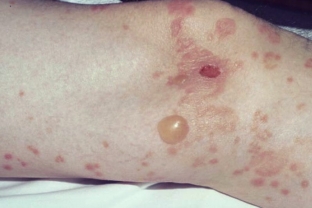
- Bullous scleroderma is accompanied by the appearance of foci of skin thickening, its sclerosis, often accompanied by hemorrhages.
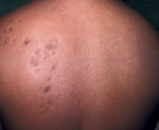
- Knotty (keloid-like) scleroderma is accompanied by the formation of multiple or single nodes on the skin that look like keloid scars. Lesions are hyperpigmented or flesh-colored, located on the neck, trunk and upper limbs.
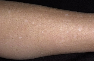
- Guttate scleroderma appears as small patches that are often associated with lichen sclerosus.
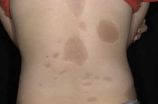
- Pasini's idiopathic atrophoderma – Pierini is considered an abortive variant of localized scleroderma. It is manifested by foci of brown color, which sink down and exist for a long time. Such foci are localized on the upper limbs and trunk, often symmetrically.
Varieties and manifestations of generalized forms of scleroderma
Characterized by the appearance of a large number of erythema foci and skin indurations that occupy several areas of the body at once, merging into extensive lesions.
Pansclerotic scleroderma is the rarest and most extensive form of scleroderma.
This form is the most severe, as it is accompanied by the formation of joint contractures with deformity of the limbs, as well as long-term ulcers.
Progressive sclerosis develops in the form of a shell, as a result, this is manifested by contracture and restriction of respiratory movements.
Eosinophilic fasciitis (Schulman's syndrome) is characterized by the appearance of painful hardening of the proximal extremities. Usually appears after injury. Sclerosis extends to deep fascia and tendons and may be complicated by carpal tunnel syndrome.
Linear forms of scleroderma are predominantly found in children and are divided into 2 types – linear and scleroderma of the "saber strike" type, learn more about which further on estet-portal.com. The linear form of scleroderma is characterized by the appearance of band-like foci of scleroderma and sclerosis, which are located on one half of the body. Linear scleroderma is usually unilateral and follows Blaschko's lines.
Scleroderma of the "saber strike" type manifested by the presence of an ivory focus, & nbsp; in which, after the inflammatory process, post-inflammatory hyperpigmentation develops.
In some cases, sclerotherapy affects not only the dermis, but also subcutaneously – adipose tissue. In this case, the foci do not have clear boundaries. There are such forms of deep scleroderma – subcutaneous scleroderma and deep scleroderma. With linear scleroderma, contractures and neurological changes in the form of headaches and seizures can form. Diagnosis of systemic scleroderma, as well as local, can be difficult. Read about the treatment of forms of scleroderma in our next article.
Share:


Add a comment