

Rare, but the most dangerous complication that can develop after the introduction of dermal fillers – blindness.
Vision loss is always preceded by occlusion of the central retinal artery.
This complication belongs to the category of ischemic, along with ischemia and skin necrosis, and develops most often after injections of hyaluronic acid.
For the period from 2000 to 2018. 53 cases of blindness after filler correction have been reported in the literature, but the actual number is believed to be much higher.
In this article estet-portal.com ophthalmologist Elizabeth Hawks (
- Mechanism of occlusion of the central retinal artery after filler injections
- Clinical signs of central retinal artery occlusion
- What to do in case of CAC occlusion after filler injections
- The role of hyaluronidase in the management of central retinal artery occlusionand
Blindness can occur after the injection of dermal fillers into almost any anatomical area.
Loss of vision, regardless of the injection site, is always preceded by central retinal artery (CAR) occlusion.
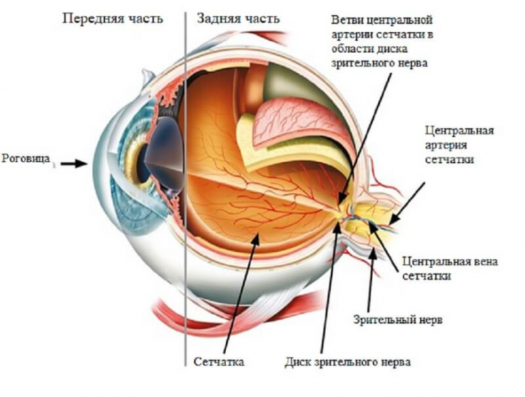
Fig. 1: Anatomy of the eye
Central retinal artery – the first branch of the ophthalmic artery, which originates from the internal carotid artery.
More details in the article: Vascular anatomy of the periorbital zone: how to reduce the risk of vision loss after the introduction of fillers
Because the central retinal artery is located deep under the skin, it is not possible to directly inject the filler into this vessel.However, the arterial network includes
branches of the ophthalmic artery connected to various parts of the face:
- supraorbital artery;
- supratrochlear artery;
- angular artery;
- dorsal nasal artery.
face areas far from the eye.
In addition, the facial artery (a branch of the external carotid artery) has several anastomoses connecting it with the blood supply to the eye.Mechanism of vision loss after filler correction
multifactorial.
The main hypothesis is that there is a possibility of direct embolization of the artery, and blindness occurs due to the movement of the embolus into the arterial network of the eye against the natural blood flow.The injecting physician must remember that even a 0.085 ml bolus of the drug can lead to dangerous consequences.
There is no clear consensus on the management of this complication.
At the same time, it is possible to prevent
permanent blindness in a short time after CAS occlusion, but in many cases, vision cannot be restored, unfortunately.
Hayreh et al. demonstrated in a primate model that retinal repair is possiblewithin 97 minutest after CAS occlusion.
Clinical signs of central retinal artery occlusionThe risk of occlusion of the central retinal artery exists when the filler is injected at
any point on the face.
But the most dangerous areas in this context are:
- nose;
- eyebrow;
- nasolabial fold;
- forehead/temples.

Fig. 2: Most often blindness develops after injections of fillers in the selected area
Sudden deterioration of vision – a clinical sign of CAS occlusion that appears immediately.
The patient may also complain about:
- pain in the eyes;
- headache;
- ophthalmoplegia;
- ptosis.
of the risk of loss of vision prior to facial fillers.
You should also pay due attention to any symptoms that occur during or immediately after injectionsWhat to do in case of CAS occlusion after filler injectionsThe Aesthetic Visual Loss Task Force (AIIVL) has published a number of guidelines containing important information for practitioners.
First step – timely diagnosis of complications based on a quick examination of the patient.
Inspection must include an assessment of:
- visual acuity;
- eye movements;
- pupil reactions.
minimum delay.
If the injector suspects an occlusion of the central retinal artery, he should immediately contact the ophthalmologist and together with the patient give him a vial ofhyaluronidase.
This tactic saves time and increases the chances ofsight recovery for the patient.
The main principle of treatment for CAC occlusion – dissolve or move the embolus to the periphery of the retina by introducing hyaluronidase into the arterial network.Hyaluronidase enzyme in cosmetology: elimination of side effects of fillers
After sudden vision loss, AIIVL recommends:
1. Injectionist:
- immediately stop infusion and contact an ophthalmologist/eye emergency department;
- ask the patient to breathe into a paper bag, which promotes vasodilation and can potentially induce clot migration to the periphery of the retina;
- prescribe a high dose of oral aspirin (in combination with gastroprotectors); as a rule, the course is 7 days;
- inject sublingual nitroglycerin to induce vasodilation and move the embolus to the periphery of the retina;
- perform eye massage;
- transfer the patient to an ophthalmologist.
2. Ophthalmologist:
- continue eye massage to stimulate clot movement (using a three-mirror lens);
- Perform anterior chamber paracentesis with a 30G needle and remove 0.1 ml of aqueous humor (in order to reduce intraocular pressure);
- reduce intraocular pressure with topical timolol 0.5% and intravenous acetazolamide (500mg);
- inject 1500 units of hyaluronidase around the bulbar area in the inferotemporal region;
- infiltrate 1500 units of hyaluronidase around the supratrochlear notch
- ki.
For any ischemic complication after
fillers, high doses of hyaluronidase (1500 units) are injected at the injection site along the arterial route.
Both an injection doctor and an ophthalmologist (depending on the situation) can administer hyaluronidase.Various methods of injecting hyaluronidase directly into the central retinal artery are under study.
Specialists review the following
injection techniques:
- retrobulbar;
- direct insertion into an artery;
- Sub-Tenon.
For occlusion of the central retinal artery, the AVIIL group recommends inferotemporal and peribulbar injection of hyaluronidase.
Hyaluronidase is able to penetrate through the walls of blood vessels, but its diffusion through the sheath of the optic nerve, where the central retinal artery lies, is impossible.
You may also be interested in: How different HA fillers respond to hyaluronidase injections: experimental results
Timely diagnosis and management of central retinal artery occlusion after filler injections is essential tominimize the risk of permanent vision loss.
According tomagazine
Share:


Add a comment