


Types of damage to biological tissues under the influence of light
Photothermal damage
Almost all dermatological laser treatments are based on heat. An increase in temperature causes the destruction of many complex molecules, which leads to coagulation of the tissue. The result depends on the method and degree of heating, from coagulation to steam generation.

Vladimir Aleksandrovich Tsepkolenko
MD, Professor, Honored Doctor of Ukraine,
President of the Ukrainian Society of Aesthetic
Medicine, General Director of the Ukrainian
Institute of Plastic Surgery
and Aesthetic Medicine" Virtus"
Thermal denaturation. At temperatures up to 43°; With even prolonged exposure does not damage the skin, from 43°; C to 50° Confirmation changes in the molecular structure begin, and tissue necrosis occurs after a few minutes. The rate of denaturation is related to the overheating temperature: its increase increases the rate of necrosis of molecules, but high temperature usually does not lead to an instant result. For example, at a temperature of 45° C human tissue fibroblasts die after 20 minutes, but withstand temperatures over 100°C for 1 ms. C. Heating the cell above 60° With at least 6 seconds leads to its irreversible destruction, and an increase in temperature by 10°; C leads to a tenfold acceleration of denaturation processes.
When a certain threshold of laser radiation power density is reached, coagulation gives way to tissue vaporization (ablation), which is an important component of laser skin resurfacing. In the process of vaporization, water molecules are superheated, turning into steam. Evaporation has a beneficial effect, since during its course most of the heat leaves the skin, but a significant increase in internal pressure leads to local "microexplosions".
If you do not stop heating after all the water has evaporated from the upper layer of the skin, its carbonization (charring) occurs, which manifests itself in the blackening of adjacent tissues and the appearance of smoke. In most cases, carbonization is a parasitic effect, leading to severe overheating of the surrounding tissues and, consequently, their extensive thermal damage.
Photoacoustic damage
With very high energy fluxes, photovaporization occurs in such a short period of time that pressure relaxation within the tissue does not have time to occur. In this case, heating leads not only to the destruction of the target area, but also to significant mechanical stresses in neighboring tissues, which causes microcracks, leading to crushing and destruction of the tissue by shock waves. Explosive processes are possible.
Mechanical damage is of great importance when removing tattoos and age spots in the process of selective photothermolysis, when high power lasers with very short pulses are used.
Photochemical damage
Under the influence of light and heat, some chemical reactions can be triggered, chemical bonds are broken, biologically active oxygen species are formed (photodynamic therapy), and cell membrane activity is increased, which improves the transport of substances. Photolysis products can change the pH of the irradiated tissue, which also activates biochemical processes.
Photochemical processes, as a rule, proceed more efficiently under the action of low-intensity ultraviolet radiation. The efficiency of visible radiation is minimal, and infrared – completely ineffective.
Instrumental diagnostics of the condition of the skin
Modern diagnostics should be based on the principles of evidence-based medicine. Subjective visual and palpatory assessment of the condition of the skin does not fully meet the above criterion. There is no doubt about the high diagnostic value of the biopsy – a method that has become the "gold standard" in dermatology. However, the invasiveness of the procedure and the possibility of scar formation at the site of biopsy sampling do not allow this method to be widely used in aesthetic medicine.
Based on the foregoing, the priority trends in the development of dermatology and cosmetology today and in the near future are becoming obvious: the development and implementation of in vivo skin examination methods.
Modern non-invasive methods for diagnosing the condition of the skin can be divided into two large groups.
1. Diagnosis of functional indicators of the skin: moisture measurement; pH-metry of the skin; assessment of sebum secretion (fat content); determination of the intensity of transepidermal water loss; assessment of skin blood flow (dopplerography); determination of melanin content and skin phototype; thermometry and skin thermography; assessment of the level of erythema.
2. Diagnosis of skin morphology: ultrasound scanning; dermatoscopy; confocal laser microscopy; optical coherence tomography; assessment of skin microrelief using reflected visible light; elastometry.
The diagnostic value of the listed methods is far from equal, and some of them need further study and improvement. The experience of many years of practice allows us to conclude that the most informative and promising are: dermatoscopy, ultrasound skin scanning, ultrasound dopplerography and remote dynamic radiation thermometry.
Comprehensive use of instrumental non-invasive methods for assessing the condition of the skin makes it possible to make a correct diagnosis, assess the severity of pathological processes in the skin, choose an adequate tactics for managing a patient during treatment, predict the likelihood of complications, the duration and effectiveness of rehabilitation measures.
Dermatoscopy is a modern method for examining the surface of the skin
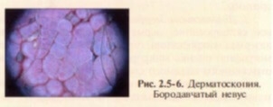
Dermatoscopy and videodermatoscopy – non-invasive diagnostic methods for visual assessment of skin lesions, allowing a more thorough examination of both the skin surface and subepidermal structures invisible to the naked eye (Fig. 2.5-6) using dermoscopic oil, which makes the surface layers more transparent. Dermoscopy is of particular importance in the diagnosis of pigmented skin lesions, which is difficult to overestimate, given the rapid increase in the incidence of malignant tumors.
Of the large number of existing methods for assessing skin neoplasms (Pehamberger Soyer, Argenziano, etc.), due to its simplicity and accessibility, the ABCD method has the largest number of supporters, which takes into account four important indicators of neoplasms: asymmetry, uneven borders, color gamut and differentiated structures . As a result of mathematical calculation of indicators, a dermatoscopic index is calculated, which is a highly informative factor in assessing skin lesions.
Ultrasound skin scan
Ultrasound (US) technologies have long established themselves as an important diagnostic tool in many areas of medicine (obstetrics, gynecology, cardiology), however, due to the insufficient resolution of sensors with frequencies below 10 MHz, ultrasound was practically not used in skin diagnostics. The development of digital imaging systems with transducers with a frequency of more than 20 MHz has made it possible to use all the advantages of high-resolution ultrasound scanning in dermatology and related medical specialties.
Modern applications of skin ultrasound include assessment of tissue edema, wound healing, imaging of skin thickness and its structural elements. This method allows you to:
determine the depth of distribution and the nature of the growth of volumetric formations, including their acoustic density, the effectiveness of the treatment of various dermatoses;
to study age-related skin changes, early diagnosis of osteoporosis, monitor the effectiveness of cosmetic procedures (external therapy, hardware cosmetology, pharmacotherapy).
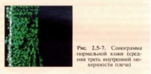
Shown in fig. 2.5-7 sonogram (acquired with a 30 MHz DUB scanner, Taberna Pro Medicum GmbH, Germany) shows a two-dimensional skin section, tissues with a lower acoustic density are displayed in a darker color. The epidermis of healthy skin looks like a thin homogeneous layer of highly echogenic tissue, and the thickness of the underlying dermis layer varies depending on the anatomical location. The dermis is visualized as a layer of fibrous structures of lower acoustic density, clearly separated from the epidermis; skin appendages and vascular elements in the form of hypo- and anechoic structures are determined in it. The subcutaneous adipose tissue located below is quite clearly delimited from the dermis and is characterized by an even lower acoustic density. With a small thickness of the hypodermis, it is also possible to visualize the muscular fascia.<
Non-invasive study of blood flow in macro- and microvessels is performed using ultrasound (US) devices based on the Doppler effect (the frequency of the signal reflected from a moving object changes in proportion to the speed of the latter). Dopplerography allows studying the blood flow both in vessels with a diameter of up to 1 mm and in arterial and venous blood vessels with a diameter of 1-7 mm, assessing the tonoelastic properties of vessels to select treatment methods, monitor effectiveness, and predict the timing of postoperative rehabilitation. The most important quantitative characteristics of blood flow are its linear and volumetric velocities, as well as the Gosling pulsation index and the Purcelo resistance index. The blood flow velocity is recorded as an integral characteristic of the tissue section.
Dynamic remote radiation thermotopography
Thermotopography, based on the capture of infrared radiation, significantly expands the possibilities of recognizing various diseases and injuries and seems to be a promising research method.
Thermotopography of the skin is determined by the characteristics of the body's heat exchange, its ability to respond to small fluctuations in the temperature of the environment, and the nature of skin vascularization. Studies show that the upper half of the human body is much warmer than the lower, and the proximal limbs are warmer than the distal. The temperature of symmetrical areas is almost similar and normally does not differ by more than 0.5 °; C. Minimal changes in skin temperature are observed in the neck and forehead, maximum – in the distal extremities.
The heat radiation of the skin depends on the central mechanisms of regulation and local factors, the main of which are the intensity of blood circulation in the skin, the level of metabolism in it and the amount of heat coming from the internal organs. Pathological conditions can affect the distribution and intensity of thermal radiation, which has both diagnostic and prognostic value (numerous clinical studies confirm this).
The diagnostic capabilities of thermal imaging have been studied in various chronic dermatoses: neurodermatitis, psoriasis, chronic lupus erythematosus, allergic dermatosis, mycoses of the feet, etc.
Thermotopography allows to determine the local change in the temperature reaction corresponding to the lesions, and to clarify the degree of activity of the process on the skin, and when observed in the dynamics – determine the effectiveness of the treatment.
Benign neoplasms of the skin are also accompanied by metabolic and hemodynamic changes and cause changes in the thermal balance, the nature of which (in combination with other methods) can be used for diagnostic purposes.
To be continued.
You can see the previous part of the article about lasers and their effects on the skin here:
How a laser works: the physical basis of the interaction of light with tissueestet-portal.com
Add a comment