


50% of all nail plate changes are fungal. Often, patients perceive it solely as an aesthetic pathology. However, onychomycosis is a long-term serious disease that, in the case of endocrine disorders, immunodeficiency, and the presence of oncology, can spread to other organs and systems. In this regard, this pathology requires timely diagnosis and full therapy. Unsuccessful treatment and further relapses undermine the patient's confidence in the reality of recovery.
- Etiology of fungal lesions of the nail plates: pathogens
- Clinical manifestations of onychomycosis: characteristics
- Dermatophytosis: topical and systemic treatment
Etiology of fungal lesions of the nail plates: pathogens
When onychomycosis affects the nail plate itself and the adjacent skin, which later is the focus of mycotic infection. The infection is spreading, sensitization of the organism by fungi and their metabolic products occurs.
Read also: Onychomycosis: diagnosis according to the latest guidelines
Pathogens are most often Dermatomycetes:
-Trichophyton rubrum;
-Trichophyton mentagrophytes;
-Trichophyton violaceum;
-Trichophyton tonsurans;
-Trichophyton schoenleinii;
-Trichophyton verrucosum.
Follow us on Facebook!
Also pathogens can be yeast fungi of the genus Candida and molds: Aspergillus, Penicillium, Cephalosporium, Alternaria, Acremonium, Fusarium ; Scitalidium.
Each type of mycosis is characterized by an individual way of penetration into the nail bed.
For example, Trichophyton rubrum first infects the skin of the proximal shaft and cuticle of the nail, and then enters the nail bed.
Clinical manifestations of onychomycosis: characteristics
The clinical picture of onychomycosis is not absolutely specific for a certain type of fungal infection. Similar symptoms can occur with various fungal etiologies.
Onycholysis – lack of communication between the nail plate and the nail bed. The separation of the nail plate starts from the free distal edge and continues towards the proximal edge. The detached part of the nail usually has a normal structure, a smooth surface, and may change color.
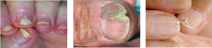
Fig.1 Oncholysis. Fig.2 Onychorrhexis. Fig.3 Onychoshisis.
Onychorrhexis – stratification of nails in the longitudinal direction. First, the furrow exfoliates at the free edge of the nail, then towards the nail matrix.
Onychoschisis – stratification of nails in the transverse direction. The nail plate retains its normal structure up to the free edge, then splits, breaks off and grows in the form of 2-3 thin plates.
Read also: Clinical forms of onychodystrophies
Median canal-like dystrophy of the nail plate looks like a deep canal-like groove 4-5 mm wide in the central part of the nail, starts from the root and moves towards the free edge, dividing it into two equal parts.
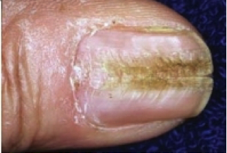
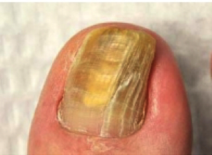
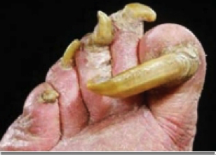
Fig.1 Median canal dystrophy. Fig.2 Onyhauksis. Fig.3 Onychogryphosis.
Onyhauksis – hypertrophy of the nail plate, which is accompanied by darkening of the nail plate. Thickening of the nail plate occurs due to the accumulation of subungual horny masses, as well as hypertrophy of the nail itself. The surface of the nail looks uneven, the color changes to gray, brown.
Onychogryphosis – thickening, curvature of the nail. The nail plate looks convex, grows upward, then continues beyond the borders of the finger. The affected nail may take the form of a horn, a spiral. The surface of the nail plate looks uneven, rough, bumpy. nail color changes to
Before deciding
with the tactics of treating onychomycosis, several factors should be taken into account:
exciter type,- matrix involvement,
- number of affected nails,
- nail plate thickness,
- Injury to the fingers or toes.
Topical treatment indicated for:
less than 30% damage to the nail plate- no or mild hyperkeratosis
- if there are contraindications to systemic therapy
, removal of the affected nail plate is carried out. After removing the nail plate, varnishes with antimycotic activity 2 – 3 bottles, these include ciclopirox, amorolfine; antifungal external agents containing ketoconazole, terbinafine, oxiconazole, miconazole – according to antimycotic susceptibility.
Systemic therapy given when:
-more than 50% of the nail plate is affected, - involvement of 2-3 nail plates in the process,
- severe hyperkeratosis, onycholysis,
- matrix damage,
- the course of the disease is more than 5 years.
As a
systemic therapyit is recommended to use systemic antimycotics – terbinafine, itraconazole, ketoconazole – according to antimycotic susceptibility. For onychomycosis caused by yeast fungi of the genus Candida, drugs containing fluconazole should be included in therapy.
Read also:Candidal onychomycosis: diagnosis and treatment The tactics of managing patients with dermatophytosis are given
according to the order of the Ministry of Health of Ukraine No. 312.Obvious is
the needfor in-depth study and determining the etiology of onychomycosis, as well as identifying the relationship with immunodeficiency, endocrine disorders and other diseases. Timely diagnosis and adequate therapy will reduce the frequency of fungal infections. Complete elimination of the pathogen and the absence of symptoms of onychodystrophy are the main criteria for completing therapy.
More interesting stuff on our