
Lichen planus (LP) is one of the most pressing problems in dermatology. This is a chronic disease of the skin and mucous membranes, characterized by the appearance of itchy, flat, polygonal reddish-pink papules with a characteristic lilac or purple tint on the skin and milky white papules on the oral mucosa. The proportion of LP is approximately 0.78-2.5% of all dermatological pathology [1, 2, 3].
Atypical forms of LP
Along with the typical manifestations of the disease, depending on the clinical picture (the nature, location and shape of the primary element), the following atypical forms of LP are distinguished: atrophic, pigmentary, follicular, erosive-ulcerative, erythematous, bullous, hyperkeratotic, staghorn, linear, annular, palmoplantar, and warty (hypertrophic) [1, 4].
The latter occurs in 15% of patients with LP. This form is characterized by papules and plaques localized on the anterior surface of the legs, with a warty pink-red surface, covered with a small number of scales. Lesions are round or oval in shape, with uneven edges and clear boundaries.
Subjectively, patients are disturbed by excruciating itching. The elements are resistant to therapy and exist for a long time. This form is extremely rarely disseminated, spreading to the skin of the trunk and extremities [1, 4].
Clinical case of warty lichen planus
We (Abdrakhmanov R.M., Khismatulina I.M., Sadykova F.G.) observed patient T., 40 years old, a resident of one of the districts of the Republic of Tatarstan. He was admitted to the Republican Clinical Dermatovenerological Dispensary with complaints of rashes on the skin of the legs (Fig. 1), back, shoulders (Fig. 2), accompanied by constant intense itching.
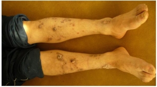
Figure 1. Pathological process on the skin of the anterior surfaces of the legs
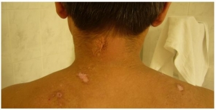
Figure 2. Pathological process on the skin of the back of the back.
Has been ill for 23 years, when elements first appeared on the skin of the outer surface of the legs. After suffering an acute myocardial infarction in February 2009, the skin process spread to the back and shoulders. In the spring of this year, he was treated on an outpatient basis at the place of residence with sedative, desensitizing, antihistamines, steroid creams with little effect.
Heredity is not burdened, does not mark professional hazards, has no bad habits.
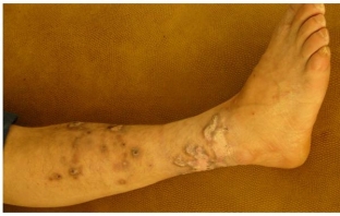
Figure 3. Pathological process on the skin of the lateral surface of the left leg.
Objectively: the pathological process is widespread, localized on the skin of the extensor surfaces of the legs, upper back, & nbsp; posterior surface of the shoulders, represented by plaques of stagnant-bluish color of polygonal shape 0.4 – 1.2 cm in diameter with a warty surface, horny and hyperkeratotic layers (Fig. 3, Fig. 4). Numerous linear excoriations.
General and biochemical blood and urine tests within the physiological norm.
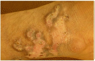
Figure 4. Pathological process on the skin of the lateral surface of the left ankle.
A histological examination was carried out, the biopsy material was taken from a site on the skin of the anterior surface of the lower leg, on the border between healthy skin and a pathological focus (Fig. 5).
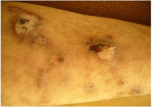
Figure 5. Biopsy site
Diffuse lamellar orthokeratosis, focal hypergranulosis, papillomatosis, uneven hyperplasia of the spinous layer (epidermal processes in some areas resemble saw teeth in shape), focal vacuolar dystrophy of basal keratinocytes, spongiosis were found. In the upper part of the dermis there is a focal and perivascular lymphohistiocytic infiltrate. Thus, the picture corresponds to that of LP.
Treatment included general complex therapy: suprastin solution 1% - 1.0 IM No. 10 and calcium gluconate solution 10% - 5.0 IM No. 10 daily, dragee «Karsil» 1 tablet 3 times a day for 10 days. External treatment was carried out by successive application with an interval of two hours of zinc ointment and the drug "Sinaflan" on previously cleansed skin twice a day for 14 days. Over the next two weeks, Naftaderm ointment was applied. 2 times a day.
In the course of treatment, positive dynamics was observed – flattening and smoothing of plaques, a decrease in itching was subjectively noted. The rash resolved within 26 days, leaving hyperpigmented spots in the lesions.
The described clinical case demonstrates a disseminated lesion that is rare in the warty form of LP, affecting, in addition to the outer surfaces of the legs, the upper back and shoulders. Thus, our observation of the atypical form of LP clearly shows the features of the manifestations of this disease, which are difficult and of interest to specialists in terms of diagnosis.
Literature
1. Gadzhimuradov M.N., Gunaeva A.A. Atypical forms of lichen planus: clinical manifestations, differential diagnosis and treatment. Clinical dermatology and venereology, №3, 2009, pp.85-80.
2. Korsunskaya I.M., Nevozinskaya Z.I., Zakharova A.B., Konstantinov E.M., Andryushkova Yu.A. Experience in the treatment of lichen planus with Glutoxim. Russian Journal of Skin and Venereal Diseases No. 1, 2008, pp. 44-46.
3. Minullin I.K., Nikitina G.V. Experience in the treatment of warty lichen planus. Practical medicine №4 (13) 2005 S. 9.4. Reference book on skin and venereal diseases for general practitioners Dyadkin V.Yu. – Kazan: Medliterature, 2006. – 320 p.
According to WWW.MEDLINE.RU, VOLUME 11, DERMATOLOGY, MARCH 2010
Share:


Add a comment