Localized (limited, focal) scleroderma is a chronic progressive disease of the connective tissue, which is accompanied by a specific lesion of the skin and underlying tissues and is characterized by the appearance of sclerotic lesions against the background of inflammation, after which atrophy and hyperpigmentation of the skin join. This disease can appear at any age and is characterized by the presence of local foci of chronic inflammation and lesions of the skin and mucous membranes with a predominance of atrophy. Recently, there has been an increase in the number of patients with focal scleroderma.
Mechanism of development and pathogenesis of focal scleroderma
Scleroderma nigricans is the most common form of scleroderma in adolescents and children.
The reason for the increase in patients with scleroderma is a change in immunoreactivity, which is associated with frequent contact with household allergens and the widespread use of antibiotic therapy.
The main link in the pathogenesis of focal scleroderma is the dysregulation of the immune system, the violation of microcirculation and the violation of the metabolism of connective tissue components. In pathology, there is a violation of collagen metabolism and defects in cellular and humoral immunity, which contributes to the formation of antibodies and the pathological process in the vascular endothelium.
Some women have microchimerism (persistence of fetal cells after pregnancy). How focal scleroderma manifests itself, read further on estet-portal.com. The fact that linear scleroderma develops along the lines of Blaschko speaks of a hereditary predisposition to the disease.
Clinical picture and complaints of patients with focal scleroderma
Patients complain of itching, soreness in the area of skin lesions, a feeling of tightness of the skin, patients note movement restrictions in the joints, as well as tissue deformation in the affected areas and changes in volume.
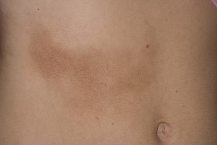
The development of focal scleroderma occurs in three stages - erythema and edema, sclerosis and skin atrophy. In most cases, the manifestation of the disease begins with the appearance of pink-lilac spots on the skin, which have a strip-like or rounded shape, sometimes swelling is present.
Further, the patches form into foci of compaction, the skin thickens and becomes ivory with a smooth surface and a waxy sheen. Skin atrophy develops with the appearance of telangiectasias, persistent hypo- or hyperpigmentation is observed.
Local scleroderma has the following course options:
- limited;
- linear;
- generalized;
- deep.
Limited form of localized scleroderma
Each variant of the course of focal scleroderma has its own characteristic and specific manifestations, as well as its own forms of the course. Thus, the limited form includes plaque scleroderma, bullous, nodular, guttate scleroderma, as well as Pasini-Pierini idiopathic atrophoderma.
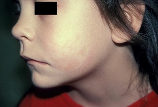
Generalized forms of focal scleroderma include pansclerotic scleroderma, eosinophilic fasciitis, saber-strike scleroderma, and progressive hemiatrophy of the face (Parry-Romberg syndrome). How each type of patchy scleroderma manifests itself, read in our next article.
Share:


Add a comment