
: строение эпидермиса")
Aesthetic medicine – This is the field of science and medicine, the main subject of study and work of which is beauty. Outer beauty of a person – this is, first of all, his skin, because it is this organ that is the mirror of our body. The skin reflects the state of all internal organs and various processes occurring in the body. Knowledge of skin anatomy is extremely important for estheticians, because it is possible to properly influence the condition of the skin only by understanding which structures the specialist works with.
With this article, estet-portal.com begins a cycle of materials about the anatomy of the skin. On the structural features of the epidermis – the outermost layer of the skin, read it now.
Features of the structure of the epidermis: basics for cosmetologists
The epidermis is the outermost layer of the human skin. It is he who is of particular interest to cosmetologists, since most cosmetic products for external application work in this layer. In addition, the epidermis – this is the first barrier faced by various damaging environmental factors that "encroach" on on the health of the human body and skin.
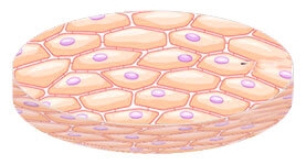
It is the epidermis that can be seen with the naked eye, so its condition is extremely important for the appearance of any person.
The structure of the epidermis is quite complex, and it is important for cosmetologists to correctly understand the anatomy and physiology of this layer of skin.
The structure of the epidermis:
- structure of the epidermis: five important layers;
- which cells provide the structure of the epidermis.
The structure of the epidermis: five important layers
The structure of the epidermis has its own characteristics that distinguish this layer of skin from others. For example, there are absolutely no blood vessels in it, and the epidermis is nourished through the dermis. The topmost layer of the skin, in turn, is divided into 5 layers:
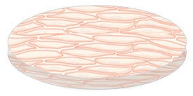
- stratum corneum is the topmost layer of human skin. It consists of several dozen rows of corneocytes – cells that do not undergo metabolism. Corneocytes contain only 10% water, are filled with keratin and do not have nuclei. They fit tightly to each other, interacting with intercellular fats, and creating a complete protective skin barrier. When the bonds between these cells are destroyed, they are exfoliated – this is how peelings work;
- shiny layer only available in areas with thick skin – palms and soles. It contains from 2 to 4 rows of flat non-nuclear cells and provides the skin with additional protection against friction;
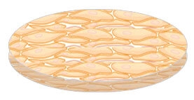
- granular layer contains up to 4 rows of small flattened cells with transparent nuclei, which are closely adjacent to each other. It is in the cells of this layer that granules of keratogeolin appear – keratin precursor. The granular layer protects the skin from dehydration and the penetration of certain substances, and also ensures the release of intercellular fats connecting the corneocytes of the stratum corneum;
-
The
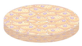
- spinous layer of the skin is so called because its cells have specific outgrowths resembling spikes. It consists of 4-7 rows of cells containing a nucleus, organelles, cytoplasm and as much as 70% water. It is in the spinous layer that the processes of keratin synthesis are launched;
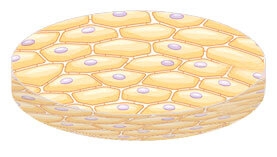
- basal layer – this is the lowest layer of the epidermis, which is the "neighbor" dermis. It has only one row of large cells, which also contain 70% water, cytoplasm, nucleus and organelles, as well as organic and inorganic substances. In the basal layer of the skin, new epidermal cells are born through active division, and then they gradually rise to the upper layers of the skin.
The granular, spiny, and basal layers of the skin are collectively referred to as the "Malpighian layer" as they are composed of living cells that have a membrane, nucleus, and cytoplasm.
Which cells provide the structure of the epidermis
The structure of the epidermis, like any other layer of the skin, is, first of all, the cells of which it consists. For cosmetologists, 5 types of epidermal cells are important:
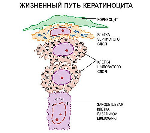
- keratinocytes – these are the most important and numerous cells of the epidermis. They are born in its basal layer and gradually move up, losing organelles and water, becoming flatter and turning into corneocytes. This process, which is called the keratinocyte life cycle, lasts approximately 26-28 days. If not at the level of the basal layer, the division of keratinocytes slows down – the thickness of the epidermis decreases, the skin looks worn and dull. It acquires the same appearance when the stratum corneum thickens and the exfoliation processes slow down – hyperkeratosis;
- melanocytes – these are the cells that produce the pigment melanin, which provides this or that skin tone. In addition, melanocytes protect the skin from the sun, which increases the production of melanin – this is skin tan. Melanocytes are found in the basal layer, but have processes that penetrate into the granular and spiny layers;
- – it is the link between all layers of the skin. They are located in the spinous layer, but with their processes they penetrate into all layers of the epidermis and into the dermis. These are immune cells that protect the skin from harmful external influences and regulate the processes of cell division in the basal layer;
- Merkel cells
- – these are receptor cells that are located in the spinous layer and are responsible for sensitivity and touch;
- stem cells
- – building material for all tissues and organs of the human body, also present in the basal layer.
protective functions of the skinFor example, timely exfoliation of the stratum corneum cells will provide a healthy and radiant appearance of the skin, and maintaining the processes of keratinocyte division at the optimal level – this is the key to the effective implementation of the
See also:
ABC of the skin for a beautician (Part 2): structure of the dermisABC of the skin for a beautician (Part 3): structure of the hypodermis Share:


Add a comment