

Tularemia – acute natural zoonotic focal disease characterized by the regular development of regional lymphadenitis and polymorphism of other clinical manifestations. Disease etiology: microorganism Francisella tularensis.
Tularemia is especially susceptible to the development of hunters due to the nature of their work and the risk of contact with infected animals, especially lagomorphs (one of the outdated names for tularemia is “rabbit plague”).
For more details about the clinical picture of tularemia, as well as the principles of diagnosis and treatment of the disease, read on estet-portal.com in this article.
Epidemiology of tularemia: main routes of transmission
The main reservoir of infection in nature – hares and rodents. The infection can be transmitted to humans through:
1. mosquitoes (main transmission vector in Finland);
2. blood-sucking arthropods (gamasid mites, fleas);
3. sick animals (during carcass processing);
4. inhalation of contaminated aerosols;
5. consumption of contaminated water or food, including the meat of a sick animal;
Thus, the following transmission routes of tularemia should be distinguished:
1. transmission;
2. contact;
3. air dusty;
4. alimentary.
It should be noted that tularemia is not transmitted from person to person.
Follow us on Facebook
Classification of tularemia: current understanding of clinical forms
The incubation period for tularemia is 1-14 days.
The development of a certain clinical form of tularemia depends on the place where the pathogen enters the human body.
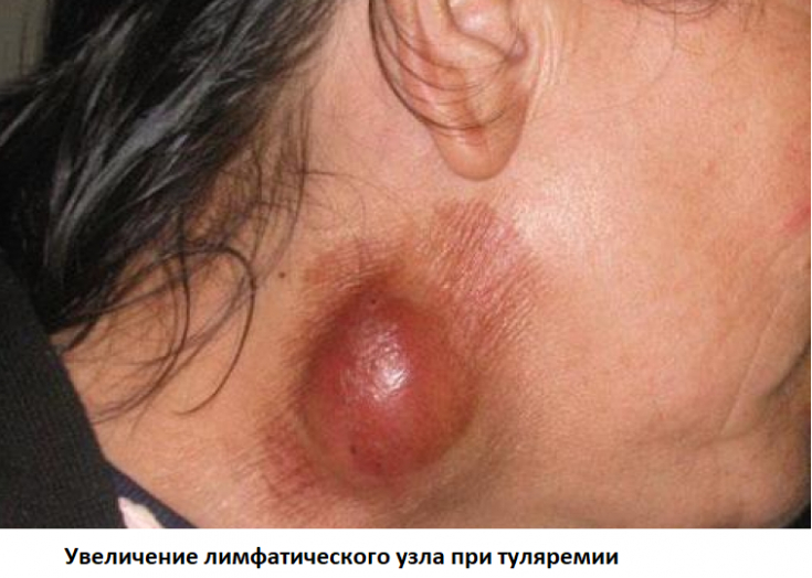
Common symptoms, characteristic of all forms of the disease, include fever, chills, lymph nodes (the latter are painful, prone to suppuration, but never solder with neighboring tissues and with each other).
Today, the following clinical forms of tularemia are distinguished:
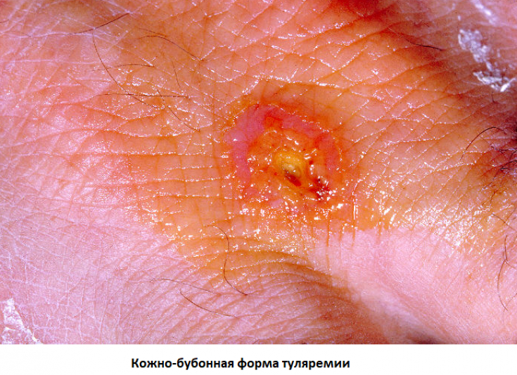
1. Dermal bubonic form: characterized by the development of a skin affect at the site of penetration of the pathogen (an itchy spot that turns into an ulcer within three days) and regional lymphadenitis;
2. Ophthalmic form: characterized by the development of mucus, photophobia, hyperemia and edema of the conjunctiva of the eyes (the process is often unilateral, but can be bilateral). Typically, the formation of yellow papules in the form of grains on the conjunctiva (granulomatous conjunctivitis), an increase in regional lymph nodes. Possible complications: dacryocystitis and corneal ulcers leading to loss of vision;
3. Anginous-bubonic form: the disease begins with the onset of pain when swallowing. On examination, tonsils are found covered with films that are not soldered to the underlying tissues. Sometimes they can develop deep enough ulcers that heal with scarring;
4. Pulmonary form: develops with airborne dust infection with tularemia, characterized by the formation of primary tularemia pneumonia involving paratracheal, peribronchial lymph nodes;
5. Abdominal (typhoid-like) form: characterized by the development of ulcers of the intestinal mucosa and an increase in mesenteric lymph nodes;
The human lymphatic system: guarding the whole organism
6. Septic form: it is very difficult and develops in people with immunodeficiency. The formation of a primary focus for this form of the disease is not typical due to the inability of the body to limit the infection. The liver, spleen, heart, bone marrow are affected. Generalized lymphadenopathy is characteristic.
Peritonitis, osteomyelitis and meningitis develop against the background of tularemia quite rarely, but such cases are still described.
Modern principles for the specific diagnosis of tularemia
The bacteriological and biological diagnosis of tularemia is associated with a large number of technical difficulties and the possible risk of infection of medical personnel, therefore, it should be carried out only in special laboratories.
Serological tests (SHA) can confirm the diagnosis of tularemia retrospectively, based on a four-fold increase in antibody titer in blood taken at two-week intervals.
Today, PCR diagnostics of tularemia is also available. For the study, samples of the separated skin affect, the contents of the lymph nodes, sputum, smears from the conjunctiva, from the oropharynx, feces and blood are used.
Primary syphilis: attention to lymph nodes
Etiotropic treatment of tularemia: effective antibiotic regimens
The treatment of tularemia is prescribed based on the presence of the clinical picture of the disease.
The emphasis in the etiotropic treatment of tularemia has changed somewhat over the past ten years.
Where previously it was thought that streptomycin was the antibiotic of choice in the treatment of tularemia, today, according to the EBM Guidelines, fluoroquinolones, in particular ciprofloxacin, are preferred.
Thus, the treatment regimen for tularemia in adults is to prescribe ciprofloxacin at a dose of 500 mg 2 times a day orally for 10 days.
Alternatively, doxycycline (100 mg twice daily for 14 days), streptomycin (500 mg twice daily IM for 10 days), or aminoglycosides may be given.
Aminoglycosides are the antibiotics of choice in pregnant women who contracted tularemia.
Thank you for staying with estet-portal.com. Read other interesting articles in the "Infectology" section. You may also be interested in When the risk of developing inflammation of the parotid gland increases
Share:


Add a comment