
Fibroma of the skin usually worries older patients, since its most unpleasant forms from a cosmetic point of view – in the form of soft wrinkled outgrowths – are formed in older women on the eyelids, on the front of the neck. Fibromas are also treated by young patients who are worried about an unexpected subcutaneous thickening. Normally, skin fibromas are harmless and easily treatable, but their histological analysis should be done in order not to miss more dangerous skin tumors with similar external symptoms.
Skin fibroma is a benign neoplasm that consists of fibroblasts and connective tissue fibers. It develops under the epidermis in the thickness of the dermis, the reasons for their appearance do not have exact explanations, a significant role of hereditary factors in the formation of fibromas is assumed. Fibromas are usually characteristic of adult patients, and their number may increase with age.
The main symptoms of skin fibroma and its varieties
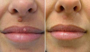
Skin fibroma usually looks like a single subcutaneous nodule, dense, well-defined, no more than a few centimeters in diameter. As a rule, fibroma does not have its own color and is covered on top with unchanged skin. But if it is large enough, it can pigment the skin a little or give it a reddish-bluish tint.
Most often fibroma has a dense elastic consistency, but sometimes there are also soft formations.
Hard fibroma of the skin (dermatofibroma) - as a rule, it is located on a wide base, but sometimes it has a so-called stalk, it can occur both on the skin and on the mucous membranes, it can be both single and multiple. To the touch is dense, painless. Such a fibroma is characterized by a "dimple symptom": if you squeeze the formation between the index and thumb, it will sink deep into the skin.
Soft skin fibroma – it looks like a wrinkled pouch on a leg, it is quite small, in size it does not exceed the size of a cherry stone of normal skin color, although it can acquire a brown tint. More often affects older women, its typical location – eyelids, front of the neck, underbust creases, armpits, groin creases.
Skin fibroma diagnosis and possible complications
The most important thing in the diagnosis of skin fibroma – watch out for dangerous tumors that may initially look similar to fibroids. Fibroma is usually differentiated from lipoma, hygroma and atheroma. When removing, it is imperative to obtain the results of a histological examination of the formation in order to finally make a diagnosis. Malignancy is not characteristic of fibromas.
Fibroma complications can be caused by its traumatization – clothes, razors. In this case, swelling, bleeding, pain are possible. If the fibroma is on the leg – there may be twisting and further necrosis. It is also dangerous to attach a secondary infection to an injured fibroma.
Treatment of skin fibroma and prognosis of its recurrence
Skin fibroma can only be treated surgically. Typically, a fibroid is left untouched if it is not a cause for concern, and removed if it is permanently injured or appears to be a clear cosmetic defect.
 Fibroma is removed under local anesthesia – either by traditional surgical excision, or with a laser or radio wave method. Today, specialists prefer laser and radio wave methods of fibroma treatment, since this involves the simultaneous sealing of small blood vessels during the incision. In addition, the surgical wound is immediately sterilized by the laser beam itself or by a radio wave. After treatment with a beam, the surface of the wound is covered with a thin layer of fibrin, which protects it from infection and prevents the development of inflammatory complications. As a result, the postoperative wound heals faster, and, as a rule, there are no traces of the operation.
With proper and timely treatment of fibroids, as a rule, in 95% of cases a complete cure occurs. After laser and radio wave removal of skin fibroids, relapses of this tumor usually do not occur.
Share:


Add a comment