

Ad oculus diagnosis in dermatovenereology still exists today. But modernity dictates to dermatologists the use in their practice of all instrumental and hardware techniques and methods to confirm the diagnosis, allowing to determine the type, and, most importantly, the cause of the disease. And early diagnosis often becomes a decisive factor in the treatment of the patient.
Find out in this article on estet-portal.com how the main diagnostic methods in dermatology can greatly facilitate the work of the doctor and increase the effectiveness of treatment.
- Histological diagnostic methods in dermatology
- Dermatoscopy – one of the most important diagnostic methods in dermatology
- Ultrasound: a promising diagnostic method in a dermatologistii
Histological diagnostic methods in dermatology
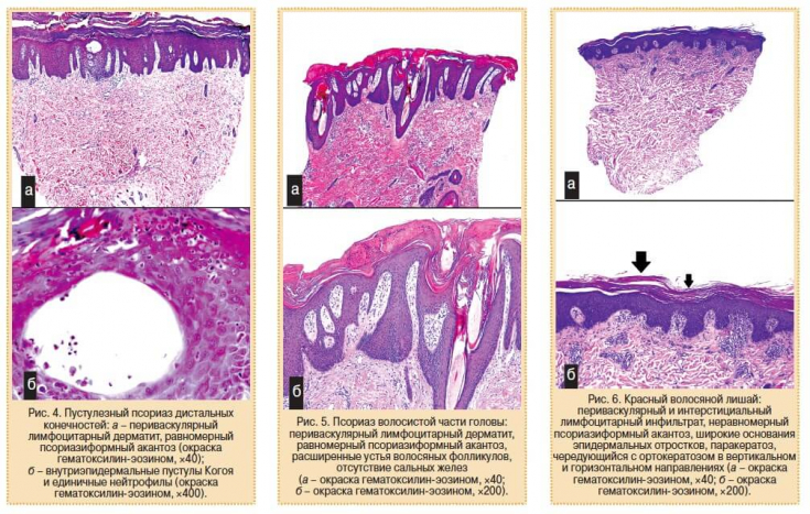
Skin biopsy is required for histological confirmation of the diagnosis. It is widely used all over the world for diagnosing not only malignant and benign neoplasms, but also for diagnosing complex and chronic dermatoses.
Follow us on Instagram!
The most common reasons for a biopsy are:
- diagnosis or confirmation;
- determination of the stage of the tumor;
- follow-up.
Types of biopsies are divided depending on the instruments they are performed, depth of intervention, tissue that is studied.
Main types of biopsies: excision, incision, perforation, shaver, curettage.
As an auxiliary method, it is possible to use direct immunofluorescent analysis, which is most often used to diagnose skin manifestations of lupus erythematosus (biopsy of the center of the lesion) and vesicular diseases, including examination of the adjacent unaffected area of the skin in case of suspected dermatitisDühring's disease, bullous pemphigoid, pemphigus.
Dermatoscopy – one of the most important diagnostic methods in dermatology
The use of dermatoscopy is gaining momentum today. This method of research of skin formations excludes surgical intervention in the human body. At the same time, a visual assessment of numerous morphological features that cannot be seen with the naked eye is carried out, which contributes to the establishment of a clinical diagnosis of almost all pigmented formations on the skin surface.
What skin changes occur with hereditary hemochromatosis?
This diagnostic method allows to improve and increase the accuracy of in vivo diagnostics, to differentiate between benign and malignant pigmented skin lesions.
Electronic dermatoscopy, using scientific methods of measuring computer diagnostics, allows to see the changes occurring in the skin, make a correct or incorrect diagnosis in a matter of minutes.
The use of dermatoscopy makes it possible to record primary skin changes in the computer's memory, monitor the effectiveness of treatment in dynamics, discuss the results with the patient, and, in unclear diagnostic cases, consult remotely with other specialists. Knowledge of the existence of these diagnostic methods not only broadens the horizons of the doctor, but also helps to refer patients with complex pathology to correct diagnosticresearch.
Ultrasound: a promising diagnostic method in dermatology
Ultrasound (ultrasound) − one of the "classic" diagnostic methods that have been used for many years in medical and research centers.
For skin examinations, ultrasound waves with a frequency of 7.5 to 100 MHz are used. Modern computer technologies make it possible to construct two- and three-dimensional images of the scanned area.
Diagnosis and treatment of actinic keratosis
Today, ultrasound technologies are actively used to monitor skin edema and wound healing, to study the structure of the skin in diseases such as psoriasis, scleroderma, and to detect tumor formations (melanoma, basal cell carcinoma).
Direct optical scanning, PRIMOS, takes a topographic picture of the skin surface and evaluates the relief from the resulting image, for example, determines the degree of roughness, "digitizes" wrinkles, scars, and the like. Another example of a direct scan system − "SIAScope" is an advanced method of dermatoscopy.
What a beautician needs to know about ophthalmic rosacea
SIAScope obtains information about the condition of the skin based on the spectral analysis of light reflected from the surface of the skin. Interest in bioengineering methods for diagnosing and examining the skin is extremely large all over the world.
Clinics, pharmaceutical and cosmetic companies, scientific institutes are interested in fast, accurate, convenient, non-traumatic methods. Today, skin bioengineering is not just an auxiliary direction, but a separate industry that successfully works in the name of human health.
More interesting information on our YouTube channel:
Share:


Add a comment