Uterine fibroids are a widespread disease. It is not uncommon for fibroids to reach gigantic sizes. Thanks to the successes of modern medicine and the increased level of culture of the population in developed countries, patients seek medical help without waiting for the tumor to become significant [1-4]. At present, despite the improvement of methods of examination and treatment, in world surgical practice, large and gigantic tumor sizes are often combined with pregnancy and are not casuistry [5, 6]. It is also known that during pregnancy, fibroids grow rapidly, reaching large sizes, and lead to disruption of vital body functions, compression of adjacent organs [7, 8]. If this tumor is combined with pregnancy, in the overwhelming majority of cases, the issue of preserving the reproductive organ is problematic, a radical operation is performed, and all the efforts of surgeons are directed to saving the patient's life. However, the experience of our clinic has shown that the preservation of not only the uterus, but also the fetus, as well as further gestation and delivery is not a myth, but a reality [9, 10]. An example of such a case is the story of patient T.
Clinical case: Patient T., 31 years old, was admitted to the obstetric clinic of MONIIAG in January 2015 with a diagnosis of giant uterine fibroids, pregnancy 13–14 weeks.
From the anamnesis: the first pregnancy in 2009 ended in urgent spontaneous delivery without complications. Small uterine fibroids were first identified in 2013, 6 months before. before the onset of pregnancy, the diameter of the largest myomatous node was 5 cm. This pregnancy was the second in the patient, it occurred spontaneously, from the early stages was accompanied by the phenomena of a threatened interruption and the rapid growth of the tumor. There were complaints of pain in the right side of the abdomen, epigastric region, shortness of breath in the sitting position. New service rewinding of electric motors For a period of 13-14 weeks. pregnancy consultatively examined at the MONIIAG polyclinic.
On examination: the abdomen is enlarged in volume according to the term of full-term pregnancy. The uterus is displaced to the left half of the pelvis, corresponds to 13-14 weeks. pregnancy. The entire right half of the pelvis and the abdominal cavity up to the hypochondrium is performed by the fibroid node, motionless, sensitive to palpation. Ultrasound examination revealed an intraligamentary uterine myoma of gigantic size (32x18x14 cm) with a pronouncedly heterogeneous structure and preserved blood flow, the placenta and the live fetus, corresponding to the gestational age, are in the projection of the tumor, the distance to the uterine cavity is from 0.9 to 1.3 cm. Surgery is recommended.
A complete clinical and laboratory examination was carried out at the clinic, which revealed Gardnerella vaginalis, Ureaplasma urealyticum, chronic herpetic and cytomegalovirus infection, anemia of moderate severity (79 g/l). Within a week, in order to prepare for surgery and prolong pregnancy, antispasmodic, antianemic therapy was carried out, drugs were administered that improve the function of the fetoplacental complex. At 14 weeks gestation. in the gynecological clinic of MONIIAG, a lower median abdominotomy was performed. During the operation, it was found that an intraligamentary cervical-isthmus myomatous node of gigantic size 32x18x14 cm originated from the region of the right uterine rib, was located completely retroperitoneally, soft, with edema and dilated vessels (Fig. 1). The body of the uterus with the fetus was pushed back by the knot to the left. Adnexa and the round uterine ligament on the right were spread on the tumor, there was a pronounced varicose veins of the small pelvis. Produced myomectomy without opening the uterine cavity with the preservation of the fetus, the node bed is sewn up with 2 rows of separate Vicryl sutures. Blood loss was 150 ml. The cavity of the right parametrium and small pelvis is drained by active drainage. Histological examination confirmed the diagnosis of leiomyoma with edema (Fig. 2). 1 dose of red blood cells and 2 doses of fresh frozen plasma were administered intraoperatively.
The cavity of the right parametrium and small pelvis is drained by active drainage. Histological examination confirmed the diagnosis of leiomyoma with edema (Fig. 2). 1 dose of red blood cells and 2 doses of fresh frozen plasma were administered intraoperatively. The cavity of the right parametrium and small pelvis is drained by active drainage. Histological examination confirmed the diagnosis of leiomyoma with edema (Fig. 2). 1 dose of red blood cells and 2 doses of fresh frozen plasma were administered intraoperatively.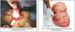
5 days after surgery, a follow-up ultrasound examination confirmed a progressive uterine pregnancy for a period of 15 weeks, the suture area after myomectomy was free of pathological formations and hematomas, there were no hemodynamic disturbances, there were no signs of a threatened abortion. Subsequently, therapy aimed at maintaining pregnancy was continued, the hemoglobin level at discharge was 91 g/l. The patient was discharged on the 10th day after the operation under the supervision of an obstetrician-gynecologist.
Thus, in order to determine the indications for myomectomy at the stage of pregnancy planning, patients with uterine myoma should undergo thorough preconception preparation, including ultrasound examination of the pelvic organs with dynamic Dopplerometry, in order to clarify the localization, size of the fibroid node and predict its growth.
Reconstructive plastic surgery for uterine myoma during pregnancy can be performed by a highly qualified gynecologist surgeon with the obligatory observance of surgical technology, which makes it possible to continue carrying the pregnancy and contributes to a safe delivery.
Literature- Buyanova S.N., Logutova L.S., Shchukina N.A. Prognosis of the growth of myomatous nodes during pregnancy (clinical-morphological and ultrasound criteria): informational and methodological letter. M.: MAKS Press, 2012. 25 p.
- Buyanova S.N., Yudina N.V., Gukasyan S.A., Mgeliashvili M.V. Modern aspects of the growth of uterine fibroids // Russian Bulletin of an obstetrician-gynecologist. 2012. V.12. No. 4. P. 42–48.
- Nappi L., Matteo M., Giardina S. et al. Management of uterine giant myoma // Arch. Gynecol. obs. 2008 Vol. 278(1). P. 61–63.
- Costa Benavente L., Silva Barroso F., Avila Flores E. Giant uterine myoma // Ginecol. obstet. Mex. 2005 Vol. 73(10). P. 563–565.
- Buyanova S.N., Logutova L.S., Gukasyan S.A., Yudina N.V. Myomectomy during pregnancy - a recognized need // Proceedings of the VI Russian Forum "Mother and Child". M., 2012. S. 20–21.
- Euzebus Chinonye Ezugwu, Chukwuemeka Anthony Iyoke, Frank Okechukwu Ezugwu, George Ugwu. Successful Pregnancy Following Myomectomy for Giant Uterine Fibroid in an Infertile Woman // J. Reprod. Infertil. 2014. Vol. 15, No. 4. P. 233–236.
- Shchukina N.A., Buyanova S.N., Babunashvili E.L. Interrelation of the receptor status of sex steroids, vascularization and growth rate of uterine fibroids // Proceedings of the VI Russian Forum "Mother and Child". M., 2012. S. 260–261.
- Kjerulff KH, Langenberg P., Seidman JD et al. Uterine leiomyomas. Racial differences in severity, symptoms and age at diagnosis // J. Reprod. Med. 1996 Vol. 41(7). P. 483-490,1088-1091.
- Krasnopolsky V.I., Buyanova S.N., Logutova L.S., Shchukina N.A. Uterine fibroids outside and during pregnancy. M.: Clinic. FUV them. M.F. Vladimirsky, 2014. 19 p.
Share:


Add a comment