
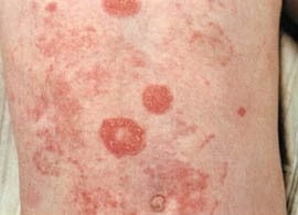
Ring-shaped skin lesions are becoming a fairly common reason for patients to visit a dermatologist, as they immediately catch the eye and cause anxiety. However, for many doctors, the diagnosis of these lesions is difficult, since symptoms such as ring-shaped or oval maculae, spots with a light center and erymatous edges are characteristic of many diseases. , and previous treatment of dermatophytosis has not been successful, other diseases with similar skin lesions should be excluded. How to understand the features of the manifestations of ring-shaped skin lesions, consider on estet-portal.com.
Some features of annular skin lesions and their comparison
We will try to present a comparison of diseases that are manifested by ring-shaped skin lesions in the form of a table.
|
Diagnosis |
Clinical signs |
Therapy |
|
Dermatophytosis of the trunk |
Inflamed, ring-shaped plaques, covered with scaly scales, located on smooth skin |
Topical and systemic antifungals |
|
Pityriasis rosea |
Oval yellowish-brown spots with small scales along the periphery, located along the lines of Langer |
Topical or systemic corticosteroids, UV treatments |
|
Granuloma annulare |
Annular plaques and papules – firm, usually not inflamed, not scaly |
Corticosteroids topically or by injection directly into the inflammation |
|
Sarcoidosis |
Erythematous plaques of a dense structure |
Corticosteroids topically or by injection into lesions, possibly antimalarial drugs |
|
Leprosy |
Annular erythematous plaques, possibly scaling |
Antimicrobial and antibacterial agents (e.g. dapsone, rifampicin) |
|
Urticaria |
Short skin lesions in the form of erymatous annular plaques without scaling |
Oral antihistamines as needed |
|
Subacute cutaneous lupus erythematosus |
Annular or psoriasiform plaques on exposed areas of the body, possibly desquamation |
Corticosteroids topically or by injection into lesions, possibly antimalarial drugs |
|
Annular centrifugal erythema |
Ring-shaped spots, erythematous on the periphery, in the center there is pityriasis peeling |
Corticosteroids topically or by injection into lesions, antihistamines |
See also: Why is it important to diagnose ring-shaped inflammatory skin lesions
Differential features of some annular skin lesions
Let's take a closer look at some of the most common annular skin lesions.
 Dermatophytosis of the trunk is a superficial fungal infection of the skin with localization on smooth skin. Elements of rashes, as a rule, are not found on the hands and feet, scalp, face and perineum. Infection usually occurs through close contact with infected persons, mainly adults are ill, although young patients are also found.
The main risk factors for dermatophytosis of the trunk – these are the characteristics of the climate (warm and humid) and the habits of people (in relation to personal hygiene).
The causative agents of dermatophytosis are promoted by a warm, humid environment, infection often occurs in showers, in pools, when using other people's towels and toiletries. When taking a history and choosing a treatment strategy, it should be remembered that long-term use of systemic corticosteroids increases the risk of developing dermatophytosis of the trunk.
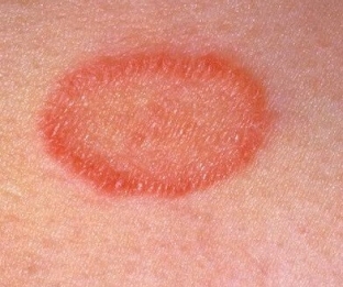
The disease presents with well-defined erythematous plaques and papules that tend to grow over time. An active inflammatory process develops along the edges of the plaques, the elements of the rash can be raised above the surface of the skin or covered with scaly scales.
The treatment of dermatophytosis of the trunk with topical and systemic antifungals is usually not difficult, but re-infection of the patient may complicate the choice of therapy. If skin lesions are localized, it is best to start with topical treatment with imidazole or allylamine preparations.
Systemic antifungals are indicated for the following conditions:
- failure or intolerance of topical therapy;
- severe disease or massive skin lesions;
- presence of chronic infections;
- immunodeficiency;
- lesion of skin areas with manifestations of hyperkeratosis.
The duration of the course of therapy is usually 2 weeks. Caution should be exercised when prescribing antifungal drugs for dermatophytosis because they can interact with other medications the patient is taking and produce unpredictable reactions.
 Pityriasis rosea is a psoriasomorphic rash that usually has a widespread sorm and often resembles dermatophytosis of the trunk in appearance.
Although the etiology of rosacea is not exactly known, current research indicates that this skin disease may be viral in nature.
Pityriasis rosea most often affects people aged 20-40 years, women get sick more often, the disease is characterized by seasonal exacerbations - in spring and summer.
Most of the elements of the rash in rosacea are macules, papules and plaques. In this case, the primary focus may look like an annular skin lesion with raised inflamed edges, finely scaly peeling and a bright spot in the center. Primary, "maternal" the focus is usually represented by an oval macula with a diameter of 2 to 10 cm. hearth may occur headaches, arthralgia, nausea and vomiting, diarrhea. Then, within 1-2 weeks, small oval yellowish-brown macules begin to appear with scaly peeling along the periphery. Elements of pink lichen are usually located symmetrically, they occur on any part of the body,
No specific treatment for rosacea is expected, but if itching is severe, topical corticosteroids and antihistamines may be given. In case of severe lichen rosacea, it may be recommended & nbsp; systemic corticosteroids and UV therapy.
Share:


Add a comment