

Scleroderma — is a chronic connective tissue disease of unknown cause. Scleroderma can be classified into localized and systemic.
Localized scleroderma affects the skin and does not affect other major organs. There are two types of localized scleroderma, namely plaque form (morphea) and localized linear scleroderma.
Systemic scleroderma involves the skin, tissues under the skin, major organs, and blood vessels. Systemic scleroderma also has 2 different types, namely localized systemic scleroderma and diffuse systemic scleroderma.
In the article estet-portal.com you can learn about localized linear scleroderma in detail
Localized linear scleroderma: onset and etiology
Localized scleroderma usually debuts between the ages of 25 and 55. However, localized linear scleroderma:
• affects children;
• more common in women;
• affects people of European descent more often than African Americans.
The onset of linear scleroderma is sometimes sudden and sometimes associated with trauma. Disease activity correlates with antihistone autoantibody titer, which decreases as the disease resolves.
Despite considerable effort, identifying the original trigger remains a significant challenge. Current hypotheses suggest a possible infectious or possibly chemical agent that activates the immune system, which in turn causes vascular damage/dysfunction and permanent fibroblast activation.
Watch the most interesting videos on our channel in Youtube
Macroscopic features of localized linear scleroderma
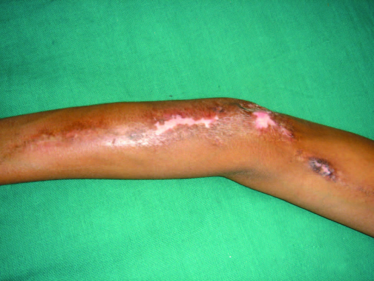
Linear scleroderma is an inflammatory disease of the dermis and subcutaneous tissue. It develops as a linear band of sclerotic hypopigmented or hyperpigmented skin, usually on the extremities. Unlike morphea, the sclerosing process in linear scleroderma can spread to the underlying dermis, subcutaneous tissue, muscles and bones and cause significant deformities.
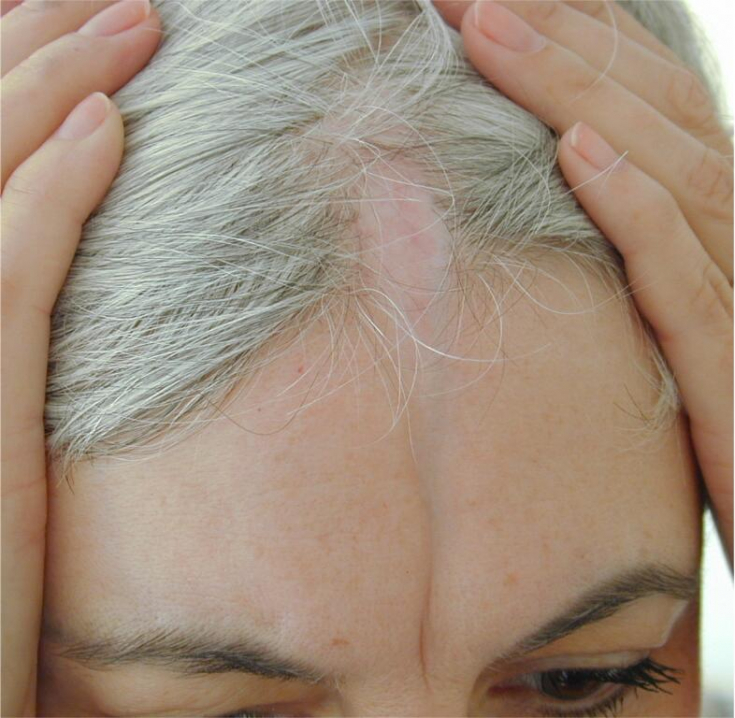
When linear scleroderma affects the face or scalp, it is called "en coup de saber"; and is associated with Parry-Romberg syndrome, a condition associated with atrophy of the hemifacial tissue below the forehead with little skin involvement.
Microscopic features of localized linear scleroderma
Localized scleroderma is characterized by three distinct features: collagen deposition in the dermis and subcutaneous layer, vascular changes, and an infiltrate of inflammatory cells, especially in the early stages.
Other histological features:
• the epidermis may be normal, somewhat atrophic, or even slightly thicker than usual;
• the dermis is enlarged and consists of wide sclerotic collagen bundles;
• collagen replaces fat around the sweat glands and spreads into the subcutaneous tissue;
• there is an increase in the number of fibroblasts;
• there is atrophy of adnexal structures, especially pilosebal units;
• eccrine sweat glands are located at a relatively high level in the dermis as a result of the accumulation of collagen underneath;
• mesenchymal elements of peripheral nerves are involved in the sclerotic process;
• vascular changes are thickening of the walls of small blood vessels and narrowing of their lumen. Fibromucinous thickening of the intima is observed in small arteries;
• The inflammatory cell infiltrate consists of lymphocytes with some macrophages and plasma cells. Immunohistochemical characterization of the infiltrate showed the presence of T-lymphocytes of both CD4 and CD8 subtypes, as well as Langerhans cells and natural killer cells.
Diagnosis of Scleroderma Buschke
Management of localized linear scleroderma
Treatment for linear scleroderma most commonly involves the use of methotrexate or antimalarial drugs, but cyclosporine and tissue allografts have also been used. The successful use of regenerative cell-enriched adipose tissue for the treatment of "en coup de saber" deformity has been reported.
A systematic review found phototherapy, methotrexate/systemic corticosteroids, calcipotriene, and topical tacrolimus to be most effective in treating morphea.
Thus, localized linear scleroderma is a manifestation of an autoimmune pathology that can be treated with modern cytostatics and hormonal drugs. Timely diagnosis is a key factor in stopping the progression process.
Thank you for staying with estet-portal.com. Read other interesting articles in the "Dermatology" section. You may be interested in Causes and clinical manifestations of focal scleroderma
Share:


Add a comment