

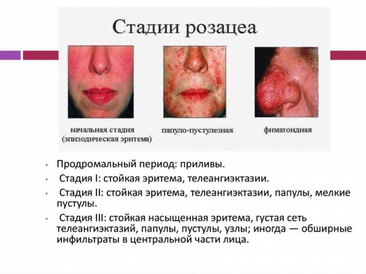
Rosacea belongs to the category of the most common dermatoses, since its population frequency reaches 1.5-10%, and in the structure of dermatological pathology - 2-8%. Rosacea is characterized by uncertainty of etiology, multifactorial pathogenesis, polymorphism of clinical manifestations, chronic relapsing course, and frequent resistance to standardized therapies. In addition, the disease causes a significant negative impact on the quality of life of patients.
Find out at estet-portal.com what are the modern methods for assessing vascular damage in rosacea.
- causes of skin and eye damage in rosacea
- Rosacea blood flow functionality
- assessment of the microvasculature in patients with rosacea
Causes of skin and eye damage in rosacea
A special place in the pathogenesis of rosacea is occupied by disturbances in the vascularization of the facial skin: the redistribution of blood flow slows down and venous stasis is formed in the area of the facial vein, which corresponds to the favorite localization of lesions.
Follow us on Instagram!
This explains frequent involvement in the pathological process of the conjunctiva, which is also located in the indicated zone of vascularization. However, at the same time, the secondary involvement of the blood and lymphatic vessels of the skin in the inflammatory process is also allowed.
Read also: Ophthalmic rosacea: when the eyes are red like a bull
It is believed that the mechanisms of vascular disorders in these patients are due to various neuroendocrine factors, which are variable, but not well understood. The available studies mainly concern the study of the morphological state of the microcirculatory bed of the skin in rosacea.
Functionality of blood flow in rosacea
The functional possibilities of blood flow, in particular, the amplitude-frequency spectrum of oscillations of the blood circulation of the dermis, remain without attention. And therefore, it seems very promising to assess the state of the microvasculature of the skin using laser Doppler flowmetry (LDF).
In favor of its use indicate:
- high information content;
- wide availability;
- non-invasiveness;
- financial affordability.
As you know, LDF is a modern technology in the field of assessing the functional state of blood circulation,
allowing you to control the features of vascularization in real time. This method is objective due to the possibility of long exposure and high sensitivity to the slightest changes in hemodynamics.
LDF is based on recording the amplitude-frequency characteristic (AFC) of a laser beam, which is reflected from blood components, mainly erythrocytes, that move in its direction.Due to the penetration of the beam into the skin to a depth of 1.5 mm, information is obtained about the blood circulation in the superficial microvessels. The particular attractiveness of this method of research lies in the fact that it makes it possible to dynamically monitor the patient during the course of the pathological process and evaluate the effectiveness of therapy.
Read also: Complex treatment of rosacea: when the skin asks for help
Tissue blood flow using LDF is usually determined in the ranges E, H, M, D and C:
- infraslow fluctuations in the range of Edue to the rhythmic activity of the capillary endothelium;
- fluctuations in the H range (vasomotion proper - rhythmic changes in the diameter of precapillary vessels) reflect the active contraction of precapillary sphincters andare under neurogenic control;
- fluctuations in the M range associated with the functioning of the ways of the juxtacapillary «shunting» circulation. Their source isthe activity of smooth muscle cells of the vessel wall and precapillary sphincters; these structural elements constantly respond to changes in intravascular pressure, that is, they ensure the implementation of the so-called myogenic reaction;
Follow us on Facebook!
- fluctuations in the range D are synchronized with the act of breathing; they are caused by an increase in blood flow to the heart at the height of inhalation and a decrease at the peak of exhalation;- fluctuations in the C range are synchronized with the heart rate and reflect changes in the diameter of arterial vessels, which are induced by the pulsation of Evaluation of the microvasculature in patients with rosacea
In a clinical study of the microvasculature of the skin in patients with rosacea, 57 patients (39 women and 18 men) aged 18 to 45 years were under observation.
The duration of the disease ranged from several months to 16 years. Was diagnosed with:
- erythematous form of dermatosis - in 22 patients;
- erythematous-papular - at 19;
- papulo-pustular - in 16 patients.
The control group consisted of 15 healthy individuals. A study was performed in the lesions in the cheek area with an apparatus for laser Doppler flowmetry. Blood flow was recorded for 20–30 min. Analysis of LDF-grams was performed using wavelet transform.
Read also: Skin blood supply: what is important for a cosmetologist to consider
Determined in the range E, H, M, D and C:- Amax (maximum oscillation amplitude)
- Fmax (maximum oscillation frequency).
It has been established that in patients with rosacea, regardless of the clinical course of the pathological process, there is a probable increase in the Amax E. This proves that the increase in the amplitude of oscillations of the capillary endothelium is combined with the inhibition of the neurogenic effect on the microcirculatory bed of the skin in the form of a decrease in the amplitude of contractions of the precapillary sphincters.
These data illustrate:
- increased activity of juxtacapillary circulation
- suppression of the amplitude of the propagation of the transmission pulsation to the venular link of the microcirculatory bed of the skin, associated with the act of breathing;
- increased tone of arterioles, stagnation of blood in the venular sections and stasis of blood circulation in the capillaries.
Somewhat different changes occurred
in the frequency spectrum of oscillations of the dermal circulation. So Fmax remained within the physiological range regardless of the clinical course of the pathological process. Therefore, the absence of possible changes indicates a discoordinated nature of the increased activity of the juxtacapillary circulation, which is due to an increase in only the amplitude of its oscillations.
Read also: Elimination of rosacea in 4 procedures. Full skin restoration
Thus, disorders of the state of the skin microcirculatory bed in patients with rosacea are branched, therefore, it is advisable for patients with rosaceato prescribe LDF studies of the skin microcirculatory bed in order to assess the depth and direction of emerging disorders, as well as amplitude- the frequency response of the state of dermal circulation should be taken into account when choosing means of therapeutic correction and can serve as one of the criteria for the effectiveness of the treatment of patients with rosacea.
More useful information on our
Share:


Add a comment