
Verrucous nevus is also called warty, because this pigmented formation is a large warty growth, outwardly resembling a cauliflower in shape. Experts attribute this nevus to the papillomatous type and consider it not dangerous in terms of the risk of developing a malignant tumor. However, if the verrucous nevus is often injured, this opens the way for infection and unpleasant complications. Therefore, in each case of such a nevus, the likelihood of its surgical removal is considered.
Distinctive features of verrucous nevus that are important for diagnosis
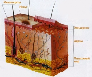
Verrucous nevus usually appears in early childhood. It grows slowly and is either a single papillomatous formation protruding above the skin, or looks like a bunch of small skin outgrowths tightly adjacent to each other. A single verrucous nevus can reach 3-4 cm in size and is most often located on the limbs, its color is flesh-colored or pigmented to varying degrees. There may be several single nevi in different areas. Systemic accumulations are usually brown in color, often arranged in garlands along the course of vessels or nerves, and can be divided into groups by unchanged areas of the skin. Such verrucous nevi are located on one half of the body.
 In childhood, a slight growth of verrucous nevi and an increase in their number is possible; in adolescents, those nevi that contain some kind of glands can grow; for example, sweat, in adults, verrucous nevi do not increase. The nevus grows not so much in width as up. Its papillomatous lobules are tightly adjacent to each other and make the surface of the nevus bumpy, rough, for which it is often called a wart. Usually such a nevus is not prone to bleeding, does not become covered with ulcers, but only if it is not injured.
Diagnostic methods confirming verrucous nevus
The specific appearance of a verrucous nevus often frightens the patient, so a thorough diagnosis must be made and the patient convinced that the nevus is melanomanohazardous. Diagnosis is a collection of anamnesis – finding out whether a nevus arose in childhood and whether its growth was observed. Further, dermatoscopy is carried out, histological examination will help to confirm the diagnosis. If the verrucous nevus is very large, located in a place where it is subject to frequent injury, or if there is doubt and a thorough histological analysis of the formation is required - – it is recommended to remove the nevus surgically.
Treatment options and prognosis for nevus verrucous
Treatment for verrucous nevus depends on its type, size, and location. Systemic nevi, occupying large surfaces of the skin with their hanging clusters, combine cryodestruction with the use of ointments containing vitamin E, vitamin A, as well as the application of salicylic vaseline compresses. Cryodestruction of such nevi is carried out in several stages and is not prescribed for small children.
In addition to cryodestruction, a good effect in the surgical excision of a verrucous nevus is shown by electrocoagulation, when it comes to the removal of single formations, as well as the radio wave method. But it should not be used for large nevus sizes, so that you do not have to suture the wound remaining after removal.
An excellent effect in the removal of verrucous nevi is shown by the laser method: excision is carried out in layers, absolutely sterile and bloodless, which is very important for large formations. But such laser evaporation of tissues does not allow their histological analysis, which must be taken into account when prescribing the procedure.
Thus, the patient needs to know that a verrucous nevus is not dangerous, but if its size is very large, it is often injured and because of this it can grow and become infected, then such a formation should be removed.
Share:


Add a comment