
In recent years, dermatologists have increasingly reminded people of the importance of constantly monitoring the condition of their skin. There are practically no people on the planet who would not have moles on their bodies. The correct name of these formations – pigmented nevi. Nevi of various sizes can appear in almost all parts of the human skin. They are a peculiar feature of appearance: the appearance of many famous personalities is remembered by people precisely because of such features. Often, pigmented nevi – these are absolutely safe formations, but under certain conditions their malignant degeneration can occur.
What can be dangerous pigmented nevi on the skin of the eyelids
Pigmented nevi are detected in one out of forty newborns, at an older age their number increases sharply, but by the age of 50 – decreases significantly. These hyperpigmented patches originate from nevus cells, epidermal or dendritic melanocytes, and fusiform melanocytes, and are local accumulations of these cells in various areas of the skin. Pigmented nevi can be located in the epidermis or in the subepithelial layer. Nevi, located on the skin of the eyelids, are potentially dangerous, as they are located in an open area of \u200b\u200bthe body and are constantly exposed to the harmful effects of environmental factors.
Pigmented nevi:
- classification of congenital and acquired pigmented nevi;
- the main types of pigmented nevi on the skin of the eyelids;
- characteristic signs of transformation of pigmented nevi.
Classification of congenital and acquired pigmented nevi
All pigmented nevi can be divided into congenital and acquired. In turn, congenital nevi are divided into three groups:
- first group - pigmented nevi up to 1.5 cm in size;
- second group – pigmented nevi ranging in size from 1.5 to 10 cm;
- third group – pigmented nevi larger than 10 cm.
Acquired nevi can be classified as follows:
- border nevi - their cells are localized between the epidermis and dermis;
- intradermal nevi – nevus cells are localized directly in the dermis;
- complex nevi – have signs of both borderline and intradermal nevi.
The main types of pigmented nevi on the skin of the eyelids
Pigmented nevi located on the skin of the eyelids can be of the following types:
- marginal pigmented nevi: often occur in childhood, are small dark spots that are located along the intermarginal edge of the eyelids;
- Juvenile nevi appear in young people, are pinkish-orange nodules that do not have a hairline on their surface;
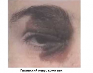
- giant nevi are found in newborns, they are large and intensely pigmented, located on symmetrical parts of the eyelids, sometimes extending to the conjunctiva;
- nevi of Ota – these are unilateral spots of red or purple color, which are located along the course of the trigeminal nerve.
Characteristic signs of transformation of pigmented nevi
The main danger of pigmented nevi of various localization, including nevi located on the skin of the eyelids, lies in their ability to malignant degeneration. Benign nevi can progress with different frequency and speed, and timely detection of signs of nevus progression determines the success of their treatment. The main signs of progression of nevi include:
- changes in the nature of pigmentation of nevi;
- formation around the nevus halo of delicate pigment;
- changes in the surface of the nevus: it becomes uneven, papillomatous;
- appearance of stagnant-plethoric vessels along the periphery of the nevus;
- increase in size of pigmented nevus.
Share:


Add a comment