

Nsolabial fold correction using traditional techniques of linear or subcutaneous injection of fillers is accompanied by the risk of various complications: hematomas, vascular occlusions with subsequent tissue necrosis in the area of the nasal alae, loss of vision. Such complications arise mainly due to the peculiarities of the location of the vessels in this area.
MD-Lift – it is a trademarked protocol developed by Dr. Dalvi Humzah. The purpose of the protocol – correction of age-related anatomical changes that lead to nasolabial folds for natural results and reducing the likelihood of serious vascular complications.
What causes the appearance of nasolabial folds
Nasolabial folds appear due to the influence of several factors, skin, soft tissues, ligaments, bone tissue and gravitational influence are involved in the process of their formation. Nasolabial folds are formed due to changes in the muscle bundles that pass through this area, as well as a decrease in skin trophism.
Follow us on Facebook
Crease border – bucco-maxillary retaining ligament. Loss of the medial deep fat pad and recession of the maxilla results in medial downward displacement of the superficial nasolabial fat pad.
These changes lead to the appearance of the nasolabial fold.
Features and goals of the MD-Lift protocol for the correction of nasolabial folds
The MD-Lift protocol was developed to correct nasolabial folds without filling them directly. This approach was inspired by the observation that deep fat compartments support superficial fat pads, and loss of volume in deep compartments leads to changes in facial volume and shape.
The MD-Lift protocol for nasolabial fold correction provides a natural result and allows you to maintain normal dynamic mobility of the target area.
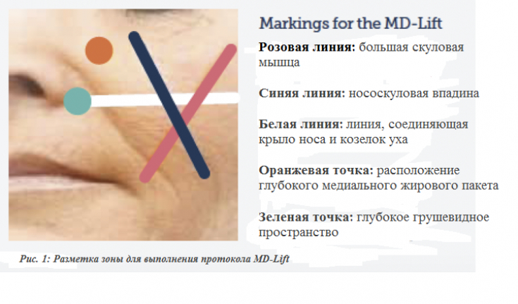
Within the framework of the MD-Lift protocol, the doctor works with two structures – deep medial fat compartment and deep pear-shaped space (Ristow's space). The angular artery runs in the superficial compartment.
Thus, injection into the preperiosteal layer or into the deep medial fat pad is relatively safe and associated with a low risk of vascular injury. In the superficial layer covering the deep pear-shaped space, the alar artery also passes, therefore, it is necessary to inject the filler in this area deeply.
Performing injection correction of nasolabial folds according to the MD-Lift protocol
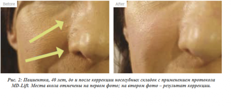
The doctor evaluates the degree of age-related changes using an approved system, takes a "before" photo, obtains the consent of the patient. After that, the skin must be carefully treated with an antiseptic. The esthetician must follow the rules of asepsis, wear a medical gown and gloves.
During the procedure, keep the working area clean and avoid contamination of the cannula, for example, by touching or contact with hair.
The anatomy of the treated area must be taken into account, in particular the location of the infraorbital, angular, and alar arteries, in relation to the cannula tip.
HA Filler Properties:
For the MD-Lift protocol, a cohesive HA filler with a high modulus of elasticity G’ should be selected, as it must provide deep tissue lifting and projection, as well as the ability to maintain its position during dynamic soft tissue movement.
To reduce the risk of vascular injury, it is recommended that the HA filler be injected using a cannula (at least 25 gauge, 38 or 50 mm). The drug is injected through one point located medially in relation to the place of attachment of the large zygomatic muscle to the zygomatic bone. If necessary, before injecting the drug, you can treat the injection point with an anesthetic.
Contour plastic: injection of filler into the nasolabial folds
From the point of insertion, the cannula is directed to the preperiosteal layer towards the deep medial fat pad. It is visualized as an area limited medially by the junction of the nasal bone with the upper jaw, from above – medial angle of the eye, laterally – nasolabial cavity (lateral border of the nasolabial fat pack), from below – lines connecting the wing of the nose and the tragus of the ear. The cannula is introduced gradually and then guided along the preperiosteal layer to the target area of injection. The cannula is placed in the deep layer at the junction of the nasal bone with the upper jaw – this is an area of deep medial fat pad. The product is injected until the nasolabial fat pad moves anteriorly (the amount of product administered varies between 0.1 & ndash; 0.4 ml).<
If skin folds remain after nasolabial fold correction (often located under the alar triangle), they can be smoothed out using the intradermal superficial injection (blanching) technique: a 30-gauge needle is inserted at an angle of approximately 10 degrees, which allows intradermal injection of a cohesive polydensified matrix . This reduces the risk of vascular damage due to the location of the facial artery in this area.
Anti-age procedures: nasolabial fold contouring
After the nasolabial folds are corrected, the injection site is cleaned and covered with a polymer adhesive to reduce the risk of contamination or infection. This gives the patient the opportunity to apply make-up immediately after the procedure.The MD-Lift protocol for nasolabial fold correction provides a natural result and allows you to maintain normal dynamic mobility of the target area.
Adapted from Aesthetics
Share:


Add a comment