


It is very important to correctly and timely diagnose diseases of the hair and scalp that cause complete or partial loss of hair, leading to cosmetic defects, thereby reducing the quality of life of such patients. In modern trichology, there are all possibilities for competent assessment of the condition of the scalp and hair, which will help to effectively prevent and slow down the development of trichological pathology.
- Trichoscopy: clinical picture of pathology
- Trichogram and phototrichogram: diagnosis of alopecia, growth phases
- Biopsy and histology: characteristic features of trichological problems
Trichoscopy: clinical picture of pathology
The procedure allows you to evaluate the main trichoscopic units: diameter and structure of the hair shaft, hair follicle orifices, scalp skin vessels, perifollicular epidermis.
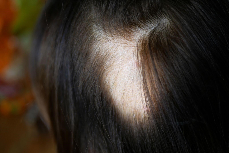
Black dots detected by trichoscopy are observed in alopecia areata, yellow dots are also found in adrogenetic alopecia.
Read also: Alopecia areata: causes and treatment
In scarring alopecia, yellow dots in the format «3D», red dots are observed occur in systemic lupus erythematosus lupus.
An important role in the trichoscopic picture is played by the features of microvascularization of the skin. So, a feature of psoriasis of the skin of the scalp is the presence of punctate or glomerular vessels. With systemic lupus erythematosus lupus in the trichoscopic picture, we can observe branched vessels inside yellow dots.
Follow us on Instagram!
During the procedure, hyperpigmentation in the form of "honeycombs" may be detected, which indicates excessive exposure to the sun; peripilar signs appearing with adrogenetic alopecia; perifollicular fibrosis characteristic of fibrous alopecia.
Read also: Trichoscopy: an effective method for diagnosing alopecia
Through trichoscopy it is possible to detect anisotrichosis - a specific sign of AGA, as well as to determine the amount of hair in follicular units, to assess their location relative to each other.
The increase and decrease of follicular units and the distance between them is a clinically important indicator for the diagnosis of androgenetic and scarring alopecia.
Trichogram and phototrichogram: features, differences
Trichogram allows you to evaluate the hair roots and the ratio of growth phases. 5 days before the procedure, the patient should be advised to refuse from washing their hair
According to the method, 60-80 hairs are depilated from two standard zones: clamping them at a distance of 0.5 cm from the scalp, they are pulled out in the direction of hair growth.
In case of suspected androgenetic alopecia, it is recommended to depilate the hair from a point 2 cm from the frontal line and 2 cm from the midline of the head. The second zone in this situation will be the area 2 cm lateral to the occiput.
The Trichogram process evaluates percentage of anagen, telogen and catagen hair. Please note that an incorrect depilation technique will lead to unreliable results.
The method is rather painful and traumatic.
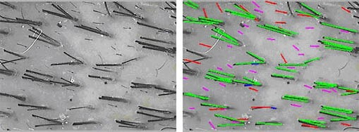
Phototrichogram is a fairly common method in trichological practice due to its accuracy and efficiency. Use both standard phototrichogram and phototrichogram with contrast.
The method allows you to study hair growth phases, determine their density and diameter, the percentage of hair in the anagen and telogen phases, the ratio of terminal and vellus-like hair, and most importantly, to evaluate the effectiveness of therapy over time.
How to influence the hair growth cyclec In the areas to be examined, the hair is shaved with a trimmer. After 2-3 days, this area is stained with an ammonia-free dye, the image is fixed at 40-60 times magnification. Next, the program analyzes the obtained data
by the number of hairs per 1 cm2 of skin, by the number of anagen, telogen, velus-like hair. Biopsy and histology: characteristic features of trichological pathology
If the
autoimmune nature of thehair loss is suspected, a biopsy with further histological examination is required. If scarring alopecia is suspected, a biopsy should be taken along the periphery of the area of hair loss; if non-scarring alopecia is suspected, – from the center of the study area.
Follow us on According to international standards, it is recommended to take 2 punch biopsies with a diameter of 4 mm each. Vertical sections are made from one biopsy, from another – horizontal. Vertical allow to evaluate changes in the epidermis-dermis junction zone, necessary for the diagnosis of cicatricial alopecia, lichenoid changes. Horizontal (transverse) sections are used to assess the total number of hair follicles, determine the ratio of terminal and vellus follicles, to detect pathological changes at the level of the upper and lower parts of the dermis, subcutaneously – adipose tissue. For a competent
trichological diagnosisyou should not stop at one method. Using a comprehensive approach, including both specialized and additional diagnostic methods, the examination of the trichological patient will be accurate and effective and will allow you to choose an individual treatment regimen.
More interesting stuff