

Practically every woman knows that, even in the absence of complaints, she should undergo a preventive examination by a gynecologist once every six months. However, a traditional examination is often not enough to reliably say whether the patient is healthy. That is why, an extremely informative diagnostic method for assessing women's health is an ultrasound examination of the pelvic organs.
But in order to correctly evaluate the results of such a study, it is necessary to remember that, depending on the phase of the menstrual cycle, a different “picture” can be seen on ultrasound. How should the ovaries and endometrium look like in different phases of the menstrual cycle on ultrasound – read today's article.
Features of ultrasound in different phases of the menstrual cycle
Every gynecologist should have knowledge of the most important physiological features of the menstrual cycle.
A healthy woman's menstrual cycle ideally lasts 28 days, with ovulation occurring on the 14th day.
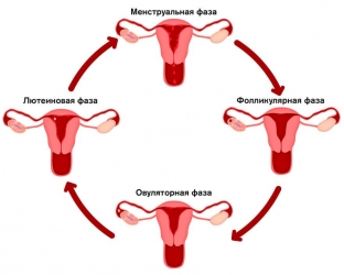
At the same time, the whole cycle consists of several successive phases: proliferation, secretion and, directly, menstruation. The proliferation and secretion phases, in turn, are divided into early, middle and late phases. In each of these phases, important physiological changes occur in the state of the endometrium and ovaries, which can be monitored using ultrasound.
But in order to correctly evaluate the data obtained on ultrasound, the doctor must know what is the norm and what – pathology for a particular phase of the menstrual cycle.
Phases of the menstrual cycle:
- what does the endometrium look like on ultrasound in different phases of the menstrual cycle;
- how the ovaries look on ultrasound in different phases of the menstrual cycle.
What does the endometrium look like on ultrasound in different phases of the menstrual cycle
Depending on which phase of the menstrual cycle the ultrasound is performed, the endometrium will be visualized differently:
- on the 5th-7th day of the menstrual cycle, in the early proliferation phase, the endometrium will have a homogeneous echostructure and relatively low echogenicity. Its thickness, on average, is 5 mm (fluctuations from 3 to 6 mm), and in the center of the M-echo, a hyperechoic thin line can be determined, which represents the border of contact between the posterior and anterior layers of the endometrium;
- on the 8th-10th day of the menstrual cycle, in the phase of average proliferation, the endometrium thickens, on average, up to 8 mm (fluctuations from 5 to 10 mm), while its echostructure practically does not change;
- on the 11th-14th day of the menstrual cycle, in the late proliferation phase, the endometrium continues to thicken, averaging 11 mm (fluctuations from 7 to 14 mm), and its echogenicity slightly increases, becoming average;
- on the 15-18th day of the cycle, in the phase of early secretion, the growth rate of the endometrium slows down, and its thickness is, on average, 12 mm (fluctuations from 10 to 16 mm). Echogenicity increases from the periphery to the center, as a result of which the hypoechoic central fragment of the endometrium takes on a drop-shaped appearance: a wide part in the region of the uterine fundus, and narrowing towards the cervix. In the phase of early secretion, the hyperechoic line in the center is not clearly visualized;
- on the 19th-23rd day of the menstrual cycle, in the phase of medium secretion, the thickness of the endometrium becomes maximum – on average, 11 mm (fluctuations from 10 to 18 mm). The echogenicity of the endometrium increases even more, and the hyperechoic line in the center is very poorly visible;
- on the 24th-27th day of the menstrual cycle, in the phase of late secretion, the thickness of the endometrium decreases somewhat, averaging 12 mm (fluctuations from 10 to 17 mm). The echogenicity of the endometrium in this period is very high, and its echostructure – heterogeneous, due to which the line of closure of the sheets of the endometrium is not visualized;
- in the phase of menstruation in the uterine cavity, a thin hyperechoic strip or hyperechoic echo structures, which are blood clots, is visualized. Sometimes the uterine cavity may look slightly enlarged due to echo-negative content, which is liquid blood.
What do the ovaries look like on ultrasound in different phases of the menstrual cycle
There are important features of visualization of the ovaries during ultrasound examination in different phases of the menstrual cycle:
- on the 5th-7th day of the menstrual cycle, in the early proliferation phase or early follicular phase, 5-10 tertiary or antral follicles are visualized, which look like round echo-negative inclusions, located mainly along the periphery of the ovary, and having a diameter of 2 to 6 mm;
- on the 8th-10th day of the cycle, in the medium proliferation phase or the middle follicular phase, a dominant follicle appears, the diameter of which is 12-15 mm, and continues to grow. At the same time, the growth of other follicles stops, and, having reached 8 to 10 mm in diameter, they undergo atresia. On ultrasound, this looks like a gradual decrease and disappearance of other follicles towards the end of the menstrual cycle;
- on the 11-14th day of the menstrual cycle, in the late proliferation phase or late follicular phase, the dominant follicle increases by 2-3 mm per day, and reaches a size of 18 to 25 mm at the time of ovulation. The fact that ovulation will occur in the coming hours is indicated by the diameter of the dominant follicle of 18 mm, the presence of a double contour around it, as well as fragmentary thickening and unevenness of the inner contour of the dominant follicle;
- on the 15-18th day of the menstrual cycle, in the early secretion phase or early luteal phase, a corpus luteum appears at the site of ovulation, the diameter of which is from 15 to 20 mm, which has an irregular shape, uneven contours, as well as a very diverse internal echostructure with varying degrees of echogenicity;
- on the 19th-23rd day of the menstrual cycle, the diameter of the «blooming» of the corpus luteum slightly increases, reaching from 25 to 27 mm, and a thickened echo-positive roller also appears. Echogenicity gradually decreases, up to the formation of a "cystic" corpus luteum;
- on the 24th-27th day of the menstrual cycle, in the late secretion phase or late luteal phase, the corpus luteum decreases in size from 10 to 15 mm ("fading" corpus luteum), its echogenicity slightly increases, and the echostructure becomes more homogeneous;
- in the phase of menstruation, the corpus luteum is often not defined, or in its place, a fuzzy echo structure of increased echogenicity remains, the diameter of which is from 2 to 5 mm. This is a white body that usually disappears without a trace during the next menstrual cycle.
There is a pronounced relationship between the anatomical and physiological characteristics of the uterus and ovaries, and the phase of the menstrual cycle.
It is the responsibility of every ultrasound technician to have the knowledge necessary to correctly interpret the resulting ultrasound image.
Only knowing how the uterus and ovaries should look on ultrasound in each phase of the menstrual cycle, the doctor will be able to determine whether the patient has a pathological condition, or all of his indicators are within the physiological norm.
See also: "Does the fight against cellulite depend on the phase of the menstrual cycle".
Share:


Add a comment