

The problem of excess body weight finds a solution in the application of techniques that allow to cause the programmed death of accumulated fat cells.
To regulate the processes of synthesis and elimination of lipocytes in adipose tissue, there is a process of apoptosis, however, it serves to remove those fat cells that have already completed their life cycle. Antioxidants are used to activate apoptosis of adipocytes, which are quite normal, but excessively accumulated.
 Maurizio Ceccarelli – biologist, professor of physiological and aesthetic medicine (Italy).
Apoptotic process in adipose tissue: the essence and features of the process
Maurizio Ceccarelli – biologist, professor of physiological and aesthetic medicine (Italy).
Apoptotic process in adipose tissue: the essence and features of the process
When a cell undergoes damage of a certain intensity, a special process called apoptosis is triggered. It leads to spontaneous cell death. An enzymatic cascade is activated, which begins with the release of cytochrome C from mitochondria.
The latter binds to pro-caspases, activating them into caspases and determining the incorporation of endonucleases. These enzymes fragment the nucleic material into a protease, then lyse the cytoplasmic material, resulting in the expression of phosphatidylserine residues outside the cell membrane.
Macrophages, not recognizing them, phagocytize as parts of cells and excrete from the body.
Apoptosis – it is a process of programmed cell death. It is characterized by a series of biochemical processes that lead to certain changes in the cell with its subsequent death.
These changes include blebbing (when bubbles appear on the membrane), nuclear fragmentation, chromatin condensation, and fragmentation of chromosomal DNA.
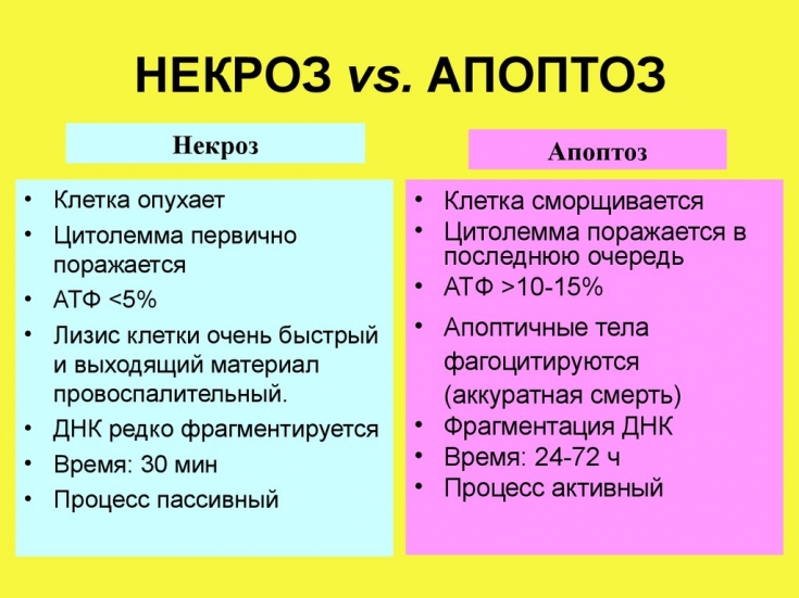
Unlike necrosis, which
is a traumatic form of cell death and occurs as a result of cell damage, apoptosis leads to cell division into apoptotic bodies that phagocytes can engulf and quickly remove without releasing intercellular substances responsible for the inflammatory process. Single cell starts intercellular
apoptoticprocess as a response to stress or, as mentioned earlier, to irreversible damage. The process is characterized by cascade activation of enzymes, controlled by regulatory proteins, which can interrupt this process at any time.
Mechanism of adipocyte apoptosis
K
apoptosisis caused by different systems, but all of them are characterized by the same intercellular mechanism. Apoptosis-inducing proteins (TNF-R1, Fas) cause mitochondrial swelling by forming pores in the membrane and increase the permeability of the mitochondrial membrane.
By increasing membrane permeability, mitochondrial proteins known as SMACs are released in the cytosol. SMACs bind to and deactivate an inhibitor of apoptosis (IAP), preventing IAP from stopping the apoptosis process, and therefore helping it to continue.
Normally, IAP inhibits the activity of a protease group called caspase. There are two types of caspase: initiator and effector. The activated effector caspase stimulates a series of intercellular proteins (endonuclease, protease) to start the cell death program.
Cytochrome C is released from the mitochondria and, once released, combines with
apoptoticprotease-1 (Apaf-1) activating factor, procaspase-9, to create a complex known as the apoptosome. This is the active form of caspase-9, which in turn activates the caspase-3 effector. Damaged during
apoptosiscells are excreted, as mentioned above, by phagocytes in a process called efferocytosis. More precisely, damage results in the release of phosphatidylserine on the surface of apoptotic bodies. Phosphatidylserine is normally located on the cytosolic surface of the plasma membrane, but during apoptosis is distributed to the extracellular surface. These molecules are the stimulus for macrophage phagocytosis. Excretion of cells by phagocytes occurs in an orderly manner, without causing an inflammatory response.
Ascorbic acid and its role in the activation of adipocyte apoptosis
process in adipose tissue serves to eliminate adipocytes that have completed their life cycle. Our goal is to activate this process and cause the death of normal lipocytes present in excess. To activate this process, we will use ascorbic acid and iron (trivalent) as a transition metal.
Studies on the activation of various antioxidant substances have shown that their antioxidant function is manifested only when these substances are present in a limited concentration.Their excess creates a paradoxical effect, leading instead of protection from damage to its activation due to the production of free radicals. This applies to all antioxidant substances, in particular,
ascorbic acid, which, in the presence of transition metals, leads to the activation of the Fenton reaction and the formation of hydroxyl radicals that damage the cell. Earlier studies have shown the ability of ascorbic acid to damage adipocytes, and its optimal concentration has been identified - 0.12-0.24% to activate the process of
apoptosis(The effect of local injection with vitamin C to adipocytes apoptosis, Gao Yan Wei Qiang Chen Shi Hai, Journal of Chinese Medicine Research, 2009 Volume 9 No. 6 pp. 325-329). Another study (Relationship between ascorbyl radical in ten sity and apoptosis-inducing activity, Sakagami H, Satoh K, Ohata H, Takahashi H, Yoshida H, Iida M, Kuribayashi N, Sakagami T, Momose K, Takeda M., Anticancer Res. 1996 Sep-Oct;16(5A):2635-44) reports induction of apoptosis with 1-10mM ascorbic acid.
With simple calculations, knowing the molecular weight of ascorbic acid (176), we can deduce the amount of vitamin C that should be used: 5.68 mmol.
Apoptosis, activated by ascorbic acid, proceeds inside the cell and is inextricably linked with the intracellular formation of hydroxyl radicals. In the presence of transition metals, in this case – ferric ion, ascorbic acid activates the Fenton reaction with the release of free radicals of the hydroxyl type. An increase in the number of free radicals leads, at the cellular level, to the activation of calcium channels with an intercellular increase in the amount of this ion. An increase in the number of calcium ions leads to an increase in mitochondrial permeability with the release of cytochrome C and cascade activation of caspase.
Thus, the process of cell death occurs after the final activation of the endonuclease, which fragments the nucleus, and the proteases divide the cell into small fragments, the so-called
apoptoticbodies, after which phosphatidylserine residues are released on the membrane surface. Phosphatidylserine residues render apoptoticbodies heterologous, stimulating phagocytosis and their subsequent digestion.
A feature of
apoptosis, in that it differs significantly from necrosis, is the absence of inflammation. The cell is engulfed by macrophages without releasing inflammatory mediators. Both apoptosis and necrosis lead to cell death, but the former is not characterized by cell destruction and the release of substances that can activate the inflammatory process. Therefore, we must be sure that the concentration of ascorbic acid, in addition to the expected result, will not cause damage to the cell membrane.
In essence, the free radicals produced in the first stage of the process, if their concentration does not exceed the permissible norm, induce
apoptosis. In the case of an excess of free radicals, they can cause the process of lipid peroxidation of the walls, damaging them and leading to cell destruction. Distinguish
apoptosisfrom necrosis using a fluorescent microscope and different stains. Apoptosis is characterized by a positive reaction to Annexin V and indicates the presence of phosphatidylserine. Necrosis characterizes positivity for propidium iodide, which indicates the presence of nuclear chromatin.
Microscopic and clinical evaluation allows us to state that the concentration of ascorbic acid 40 mg/ml is safe and induces apoptosis without necrosis.We also have to take into account another important point: the hyperosmolarity of ascorbic acid. Applying the van't Hoff rule, which relates the osmotic pressure to the molar concentration of a substance, we determined the osmolarity of the injected ascorbic acid. It is seven times the normal value.
Saline (0.9% sodium chloride) is so named because it does not affect the osmotic balance of tissues. Calculating its osmotic pressure gives us a value of 310mOsm/l.
The result comes very quickly:
apoptosis– it is a process that takes place over several hours, it is visible after 2-3 days and remains unchanged.
Succinic acid - active ingredient to combat hyperpigmentation
Share:


Add a comment