Infiltrative-suppurative trichophytosis is caused by fungi that parasitize humans and animals, that is, it belongs to the zoophilic group. Objects contaminated by animals can become a source of infection for humans. This disease often causes difficulties in diagnosis and therefore its advanced forms are often encountered.
The trichophyton species that cause disease in humans are of the ectothrix type and are located on the outer surface of the hair, forming a "sheath" around it.
Depending on the size of the spores and their biological characteristics, a small-spore variety (microides) is distinguished, which parasitizes small domestic animals, mice, rats, rabbits and guinea pigs, and a large-spore variety (megaspores) - in large domestic animals: horses, cows, calves , sheep, which is characteristic of Trichophyton verrucosum (faviforme). The skin lesion caused by them - infiltrative-suppurative (deep) trichophytosis - is characterized by the formation of a perifollicular inflammatory infiltrate, leading to purulent fusion of the hair follicles and the surrounding connective tissue.
The source of infection with infiltrative-suppurative trichophytosis are animals with this mycosis through direct contact with people (direct route) or indirectly through objects that have hair of sick animals infected with fungi.
Clinical case
Patient N., born in 1988, complained of rashes on the scalp.
Anamnesis of the disease: considers himself ill for 1.5 years, when rashes first appeared on the skin of the scalp. The patient associated the occurrence of rashes with hypothermia. In this regard, he sought medical help from a surgeon.
The diagnosis was made: "Multiple abscesses of the scalp". Surgery was performed to open abscesses, intravenous levofloxacin, vancomycin, fluconazole, and antistaphylococcal immunoglobulin were prescribed. The wounds cleared, the patient was discharged in a satisfactory condition.
At the same time, clinical manifestations resumed, in connection with which he repeatedly underwent outpatient treatment at dermatovenereologists with a diagnosis of "Undermining folliculitis of the scalp", confirmed by the detection of epidermal staphylococcus aureus during bacteriological examination. Mycological examination was not performed.
Due to the lack of effect from the treatment, he sought medical help at the Department of Dermatovenereology of the NMAPE named after P.L. Shupyk, was hospitalized in the Kyiv City Clinical Dermatovenerologic Hospital.
Objectively: on the scalp, mainly in the parietal-occipital region, there are deep, dense, sharply limited, tumor-like elevated inflammatory infiltrative foci, intensely hyperemic, with massive purulent crusts on the surface. In the central part of the foci there are fluctuation phenomena with the destruction of the skin and hair follicles. Hair falls out, a copious amount of pus is released from the holes of the empty follicles. In place of resolved foci, there are retracted scars, an abundance of purulent hemorrhagic crusts (Fig. 1).
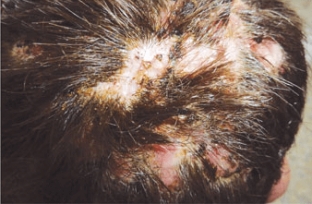
The posterior and anterior cervical lymph nodes are enlarged, painless, not soldered to the underlying tissues. An increase in body temperature to subfebrile numbers is noted.
From the epidemiological history: according to the patient, he did not come into contact with large domestic animals, although he repeatedly traveled to the countryside for a short time.
Upon admission of the patient to the hospital, a bacteriological examination of the contents of inflammatory foci, a microscopic examination of skin and hair scales were carried out.
Bacteriological culture of the contents of inflammatory foci revealed hemolytic staphylococcus aureus.
Microscopy of skin flakes revealed an abundance of mycelium and spores of the parasitic fungus, in connection with which the hair and skin flakes were sent for examination for parasitic fungi. Bacteriological examination of hair and scales revealed faviform trichophyton, the causative agent of deep trichophytosis (Fig. 2, 3).
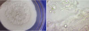
Tr. verrucosum (faviforme) - faviform trichophyton, the causative agent of deep trichophytosis. Affects the skin of the scalp, occasionally nails. Microscopically in hair it is defined as large-spore trichophyton. This type of dermatomycete is best cultivated on peptone agar. Therefore, if the presence of a pathogen is suspected, pathological material is sown in parallel on Sabouraud's medium and on peptone agar. Culture growth is observed on the 7th–20th day in the form of a gray hump, deepened into the medium; when examining a colony under magnification, on its periphery, the location of mycelium hyphae in the form of rays is determined. Microscopically: mycelium with thick septate hyphae, tips smooth or pointed.
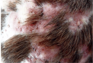
Treatment. The patient was prescribed griseofulvin according to the scheme for 3 weeks. After the treatment, the patient's condition improved significantly, body temperature returned to normal, the pathological process on the scalp is resolved, inflammation in the foci disappeared, there is no fluctuation, hair grows in the affected areas (Fig. 4).
This case demonstrates the importance of laboratory diagnostics, in particular, bacteriological examination of pathological material (skin and hair scales) when the process is localized on the scalp.
To conduct a differential diagnosis, it is necessary to determine the range of laboratory tests that will allow timely establishment of a clinical diagnosis, especially in case of suspected skin infection (bacterial, fungal, viral), and to carry out an appropriate set of anti-epidemic measures to prevent the spread of this infection.
According to kiai.com.ua
Share:


Add a comment