
Protruding dermatosarcoma is also called lumpy fibrosarcoma of the skin or recurrent dermatofibroma. This is a rare connective tissue tumor that has an average degree of malignancy. The tumor grows over several years or decades with a gradual introduction into the underlying tissues, is not prone to metastasis. More often, dermatofibrosarcoma is detected in young people. For timely detection of a tumor, it is important to know its external manifestations and the nature of growth. Read more about this in our article.
What are the causes and mechanism of development of dermatofibrosarcoma?
The causes and mechanism of development of dermatofibrosarcoma have not been fully established. There are two theories of its development - it is vascular and fibroplastic. Thus, some researchers claim a connection with trauma, while others refer to the presence of chromosomal abnormalities during the growth of dermatofibrosarcoma.
The size of the neoplasm can range from a few millimeters to 15 centimeters. The size depends on the duration of the course of the disease. Detection of dermatofibrosarcoma occurs when its size reaches several centimeters.
Dermatofibrosarcoma appears to be fibrous, dense, composed of bundles of spindle-shaped monomorphic cells. It is possible to identify atypical mitoses. Sometimes there are signs of mucosal degeneration. What the tumor looks like, read further on estet-portal.com. Fibroblast bundles and collagen fibers of dermatofibrosarcoma form a variety of structures that resemble bundles, rings and swirls, so the tissue acquires a characteristic "moire" pattern. view.
The main symptoms of the development of dermatofibrosarcoma bulging
Dermatofibrosarcoma usually appears at the age of 20-40 years. The tumor is more often localized in the back, chest, abdomen or proximal upper extremities. Rarely, the tumor appears on the scalp or neck. Dermatomyosarcoma in the early stages resembles a scar or lipoma in appearance. In most cases, a single tumor is found.
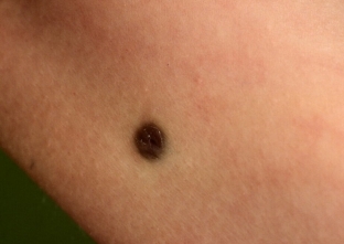
Characteristics of dermatofibrosarcoma:
- The surface of dermatofibrosarcoma is smooth or slightly bumpy, brown or red.
- The skin over the tumor is tense and atrophied.
- The tumor is initially mobile, painless.
- Erosion covered with serous – hemorrhagic crusts.
- It is important to consider that, although the tumor does not metastasize, it can grow deeply and affect the bones, fascia, muscles and organs located below them.
The purpose of diagnosis and treatment of dermatomyosarcoma
The diagnosis is made on the basis of clinical characteristic manifestations and data from additional studies. Since there are few specific symptoms in the manifestation of dermatomyosarcoma, the main diagnostic methods are histological and microscopic examination of the biopsy.
In the presence of bundles of elongated cells and signs of spindle cell proliferation, "moiré" pattern and atypical mitoses, dermatofibrosarcoma is diagnosed.
No specific immunochemical markers. The diagnosis is differentiated with tumor forms of mycosis fungoides, dermatomyosarcoma, fibrosarcoma & nbsp; gummy syphilis.
Treatment of dermatofibrosarcoma is surgical, carried out in a planned manner. The most effective method of removal of dermatofibrosarcoma according to the Mohs method, in which the tumor is excised, capturing a few centimeters of unchanged tissue.
Share:


Add a comment