Pigmentary nevus of the skin is a benign formation that is located simultaneously in the epidermal layer of the skin and the dermis. A pigmented nevus has the appearance of a papule or wart that rises above the level of the skin by about 1 cm.
This type of nevus belongs to the main types of melanocytic nevi of epidermal origin. The nevus begins its growth in the epidermis, and ends in the dermis, which distinguishes the pigmented nevus of the skin from moles. Therefore, a pigmented nevus is called a complex nevus. Read more about clinical manifestations, diagnosis and treatment of pigmented nevus.
Main clinical manifestations of pigmented nevus of the skin
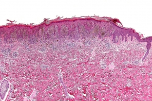
Complex pigmented nevus of the skin combines signs of intraepidermal and intradermal nevus. The intense brown color of the nevus is due to the epidermal component. Is the pigmented nevus of the skin dangerous, knows estet-portal.com. According to many researchers, up to 80% of nevi degenerate into melanoma, despite the initially benign nature. Therefore, in dermatology, pigmented nevus of the skin is classified as melanoma dangerous.
The intradermal component of the nevus contributes to its elevation above the skin surface. Therefore, it resembles a wart.
Pigmentary nevus of the skin looks like a node or papule with a smooth surface. Sometimes there are complex nevi with a warty or keratinized surface. More often, a pigmented nevus of the skin is localized on the scalp, but may have a different localization. The nevus can be up to 1 centimeter in diameter.
Diagnosis. Methods for diagnosing pigmented nevus of the skin
The diagnosis of "complex pigmented nevus of the skin" is made on the basis of examination by a dermatologist, skiascopy and dermatoscopy. In order to determine the degree of germination of the nevus in the dermis, ultrasound of the skin formation is used. If melanoma is suspected, the patient should be referred for a consultation with a dermato-oncologist.
A biopsy of a pigmented nevus of the skin is dangerous because of its possible transformation, which can provoke a malignant degeneration into melanoma. Therefore, a histological examination of the tissues of the nevus is carried out after its complete removal.
Differential diagnosis of pigmented nevus of the skin is carried out with the following pathologies:
- blue nevus;
- warts;
- papilloma;
- Setton's nevus;
- Dubreuil's melanosis;
- border pigmentary nevus;
- dermatofibroma.
Tactics of treatment of pigmented nevus of the skin
In the presence of a pigmented nevus of the skin, the patient should be under the supervision of a dermatologist. With constant trauma to the nevus, as well as at the risk of its malignancy, removal of the nevus is indicated. Also, the removal of a pigmented nevus of the skin can be done for cosmetic reasons. Removal of the nevus is performed by laser, surgical excision and radio wave method. Cryodestruction and electrocoagulation are not used due to the risk of possible trauma or incomplete removal, and also because it can stimulate malignant growth.
In any case, if a dark-colored elevation is found on the body, you should not try to get rid of it yourself. Consult a dermatologist to determine further tactics for removing a pigmented nevus on the skin.
Share:


Add a comment