

Facial Aging – an irreversible process, which is a combination of changes at the level of bone structures, fat, muscles and ligaments, as well as skin and blood vessels. All of them are interconnected. Loss of volumes and redistribution of adipose tissue – certainly an important factor that leads to age-related changes in the shape and contours of the face. To "return to place" soft tissue, in aesthetic medicine resort to filler injections. In the estet-portal.com article, Dr. André Luis Castro De Magalhães Mattos looks at the aging process, with a special focus on adipose tissue, and provides an overview of facial reshaping techniques with lipofilling and hyaluronic acid injections.
- Structural Components of Facial Aging
- Facial lipofilling to restore soft tissue volumes
- Facial correction with hyaluronic acid injections
- Possible complications after soft tissue fillers
Structural components of facial aging
With age, there is a noticeable reduction in the height of the face, which is mainly due to changes in the upper and lower jaws, as well as a slight increase in the width and depth of the face. The eye orbits increase in size while the maxilla decreases, resulting in a downward displacement of the malar fat pads and a deepening of the nasolabial fold.
The aging of the craniofacial skeleton is a consequence of atrophy and changes in the relative dynamics of growth and bone loss.
Resorption of the maxilla can also deprive the upper lip of its support, leading to the formation of perioral wrinkles. In the lower jaw, tooth loss leads to severe resorption of the alveolar process. The shape and projection of the chin also change with age. In addition, there is a thickening of the tubercles of the lower jaw at the points of attachment of the masticatory muscles (the angle of the lower jaw and the lower edge of the zygomatic eminence), as well as softening in other areas of the face.
Subscribe to our channel in Telegram!
Subcutaneous fat distribution
Young face is characterized by an even distribution of superficial and deep fat, which provides youthful curves and bulges of the face.
Facial aging is associated with loss of soft tissue volume in certain areas:
- periorbital;
- forehead;
- painting;
- temporal;
- mandibular;
- chin;
- glabellar;
- perioral;
as well as fat hypertrophy in other areas:
- submental;
- lateral part of nasolabial and labiomental folds;
- brel;
- infraorbital;
- painting.
In addition to redistribution and loss of volume, fat pads become more distinct, as do other facial structures such as the submandibular glands and bony tubercles.
With age, the malar fat pad gradually shifts forward and downward and bulges near the nasolabial fold, thereby increasing its severity. This redistribution and demarcation of fat gives the face an unbalanced appearance.
Read also: Effective approaches to restore the volume of the middle third of the face
In the periorbital and perioral areas, muscle activity results in dynamic wrinkles that become static over time.
Sagging (flaws, submental area and nasolabial fold) is formed due to excess skin and/or lack of elastic recoil, as well as fat accumulation.
As a result, the curves and bulges of the young face are flattened. The line of the lower jaw becomes uneven, depressions form in the temporal, buccal and infraorbital zones, the lips become more straight and angular. The bend of the lower jaw is smoothed out, flares appear, the forehead and eyebrows lose their anterior projection.
In order to restore a young look to the face, the author prefers to restore the volume of soft tissues through injections of hyaluronic acid and lipofilling.
Facial lipofilling to restore soft tissue volumes
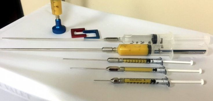
The principles of fat grafting are based on the optimal vascularization of the recipient site, which is necessary to improve the survival of adipose tissue.
Through an incision in the skin with a size corresponding to the diameter of the cannula, the fat graft is injected at the level of the target anatomical zone. It is believed that small-caliber cannulas reduce the degree of trauma to the recipient zone and thereby reduce the risk of bleeding, hematomas, and insufficient oxygen diffusion in the graft. Since revascularization starts from the periphery, the center of the graft is characterized by a longer ischemia time.
Therefore, multiple injections of small volumes are preferable to a single injection of a large volume of fat. Typically, multiple channels are created through multiple access points, however fat is injected only during cannula withdrawal in the fan technique. To avoid excessive interstitial pressure in the recipient zone and crowding of transplanted adipocytes, fat grafts are injected in small aliquots and distributed in a fan-shaped manner at different depths of the soft tissues.
Studies have shown that mobile areas of the face, such as between the eyebrows and lips, are less amenable to correction than more static areas, such as the cheekbones and lateral cheeks.
Lipofilling of various areas of the face for optimal rejuvenation and beautification
Fat injections use different sizes of cannulas, and the nature of the recipient site is the main determining factor in this matter. Fat grafting continues to be the procedure of choice for soft tissue augmentation during surgical ofacial procedures.
Facial correction with hyaluronic acid injections
Scientists and doctors are constantly looking for the perfect dermal filler, which should be:
- safe;
- effective;
- biocompatible;
- non-immunogenic;
- easy to distribute and persist in tissues;
- Easy to remove if necessary.
HA fillers have the most optimal characteristics, therefore contouring with their use rightfully ranks second among the most popular non-surgical procedures for women and third – for men.
There are various types of HA fillers available on the market for correcting moderate to deep facial wrinkles and folds, as well as for volume augmentation. The insertion technique depends on the product chosen and the goals set.
For the purposes of volumization, the author prefers to use the Redka-Galadari method (Redka-Galadari) in his practice:
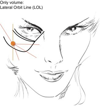
Point RG – an anatomical safe zone, which is located along a perpendicular line extending from the lateral corner of the eye to a point located 1–2 cm below the orbital ridge. From this entry point, the filler can be inserted into the cheeks and nasolabial folds, and, depending on the size of the cannula, into other areas of the face. To avoid complications, it is important to know exactly in which plane the product should be injected.
Possible complications after the introduction of soft tissue fillers
In recent years, the number of procedures using soft tissue fillers has increased significantly, and accordingly, the number of complications has also increased.
Filler complications are classified according to:
- severity (mild, moderate, severe);
- start time (early and late);
- character (ischemic and non-ischemic).
You may also be interested in: Management of early complications of contouring
The expert group proposes to categorize filler complications into:
- immediate (develops within 24 hours after the procedure);
- early (24 hours & ndash;4 weeks after the procedure);
- delayed (>4 weeks post-procedure).
The most serious complication that can develop after the introduction of any filler, – vision loss.
Vascular Occlusion – a rare but very dangerous complication that occurs due to the introduction of filler material into a blood vessel.

Fig. 1: filler in the deep plane of the temporal zone
Intravascular injection of fillers can lead to the following complications:
- ischemia and tissue necrosis;
- blindness;
- pulmonary embolization;
- stroke.
Beleznay et al. note that 47.9% of blindness cases are caused by injections of autologous fat, while for HA fillers this figure is 23.5% cases.
Autologous fat, the most viscous soft tissue filler, is associated with the highest risk of diffuse ophthalmic artery occlusion and the worst prognosis. In the case of HA, hyaluronidase can improve the outcome of complications after intravascular injection of the filler.
It is also suggested that in the case of embolization of the central retinal artery with a HA product, retrobulbar injection of large volumes of hyaluronidase may be the most effective way to quickly dissolve the filler. Important factors that ensure rapid recovery are hyaluronidase concentration and enzyme injection time.
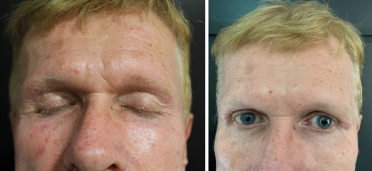
Fig. 2: before and after temporal cavity correction with HA filler
All practitioners should be prepared to respond promptly to complications following fillers. To do this, you must have a first aid kit in the office and familiarize yourself with the protocols for managing complications.
All layers of the face are involved in the aging process. Understanding age-related changes and a three-dimensional approach to face correction – guarantee of good results. Choosing the right filler for the target area is also an important factor. The risk of complications after injections of soft tissue fillers must be taken into account during each procedure, and therefore every doctor must be able to diagnose in a timely manner and quickly respond in case of serious complications after the introduction of fillers.
Adapted from The PMFA Journal.
More interesting videos on our YouTube-channel!
Share:


Add a comment