

Hematochezia (blood in the stool) is a common anorectal symptom. The cause can be inflammation, hemorrhoids, cancer, polyps, certain diseases like ulcerative colitis and Crohn's disease. One of the rare causes is intestinal endometriosis, which is always associated with difficulties in diagnosis, diagnosis and treatment of the patient.
Endometriosis originating in the digestive tract mainly affects the rectum and sigmoid colon. It most often occurs between the ages of 30 and menopause. Ectopic endometrial tissues in the rectum are usually located in the muscles and rarely in the submucosa.
Malignancy of ectopic endometrial tissues in the rectum into endometrioid adenocarcinoma is possible. Read in the estet-portal about what clinical manifestations and examination methods will help to identify intestinal endometriosis.
Description of a clinical case of intestinal endometriosis with hematochezia
A 37-year-old woman was admitted to the hospital with an anus and rectal prolapse accompanied by hematochezia. The symptoms started 3 years ago. The patient had no abdominal pain, diarrhea, or weight loss. Prolapse of the anus and rectum was detected by digital rectal examination.
Follow us on Telegram
According to these findings, the patient was diagnosed with hemorrhoids. A digital rectal examination also revealed a mass of approximately 1.5 cm.times. 1.5 cm on the rectal mucosa at a distance of 8 cm from the anus.
Laboratory and instrumental diagnostics of intestinal endometriosis
Tissue samples were examined histologically after staining with hematoxylin-eosin and immunohistochemically for the detection of steroid hormone receptors in hormone-positive tumor cells.
Specific antibodies included monoclonal antibodies to muscle-specific actin Actin (1:100, DAKO), CD10 (1:100, DAKO), CK (1:200, DAKO), Ki67 (1:100, DAKO) and estrogen receptors - ER (1: 200, DAKO).
Immunohistochemistry – method of localization of specific antigens in tissues, based on the recognition of the antigen by the corresponding antibody and the detection of the results of this binding at the light-optical level.
Computed tomography (CT) of the abdominal cavity revealed a node in the lower part of the left wall of the rectum with a diameter of 9 mm. Retroperitoneal lymph nodes were not involved in the pathological process.
Read more about what is the danger of endometriosis on estet-portal.com in the section "Gynecology".
Microscopic features of intestinal endometriosis
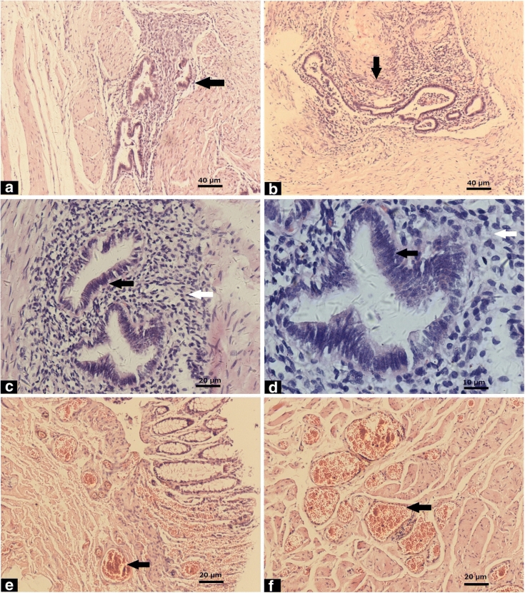
Ectopic endometrial tissues (black arrows) were mainly located in the muscles (a) and submucosa (b) of the rectum. Ectopic endometrial glands (black arrow) and surrounding interstitial tissue (white arrow) at high magnification (c, d).
Epithelial cells without severe atypia (black arrow) (D). Small vessels (black arrows) were found in the muscles (e) and submucosa (f) of the rectum.
Results of immunological studies. Actin (cm) was positive in smooth muscle cells (black arrow) and negative in the submucosa (grey arrow) of the rectum, endometrial interstitium (white arrow). CD10 was positive in the endometrial interstitium (black arrow).
CK and ER were positive in endometrial epithelial cells (black arrows). The Ki67 index in ectopic endometrial tissues was approximately 5% (black arrow).
Ki-67 - marker of tumor cell proliferative activity. Ki-67 less than 15% - the tumor is slightly aggressive, Ki-67 in the range of 30 - 50% indicates in favor of an aggressive tumor, and with Ki-67 above 50%, the tumor is highly aggressive.
Causes of unsuccessful differential diagnosis of intestinal endometriosis

Hematochezia is a common symptom of hemorrhoids and can also occur with a rare pathological process - intestinal endometriosis.
Causes of diagnostic pitfalls of endometriosis in the rectum include:
1. endometriosis in combination with hemorrhoids. The patient had a three-year history of hemorrhoids, the symptoms were fully consistent with the diagnosis.
2. The endometriosis nodule in the rectum was small and difficult to diagnose during digital rectal examination due to its submucosal location at a distant distance from the anus (8 cm).
3. Microscopic examination revealed a vessel dilatation in the intestine with endometriosis, which could be misleading and confirm the diagnosis of hemorrhoids, if extensive samples with endometrioid tissue were not taken.
Thank you for staying with estet-portal.com. Read more in the section "Gynecology" in our article Constipation in women can be a symptom of retrocervical endometriosis
Based on BMC Women's Health.
Share:


Add a comment