

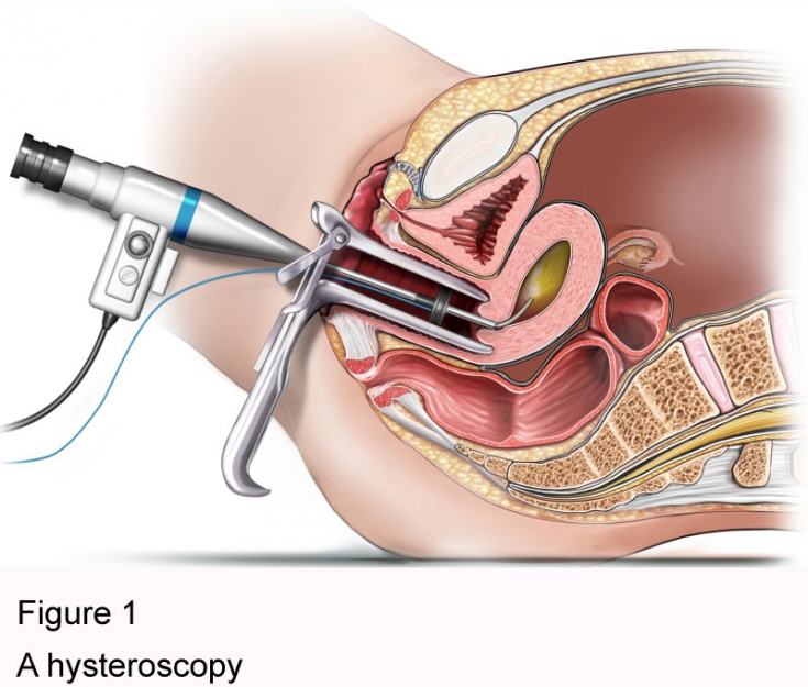
Hysteroscopy — one of the most informative diagnostic methods in gynecology, which allows you to visually assess the state of the uterine cavity and perform additional surgical manipulations under visual control. This is a microsurgical intervention that has minimal consequences for a woman's health. This method is used to diagnose and remove polyps, myomatous nodes, as well as to dissect synechiae. Read more about hysteroscopy on estet-portal.com in this article.
The main indications for the use of the hysteroscopy method
Hysteroscopy is indicated for diseases of the uterus, such as adenomyosis and tumors, and this method is also used for menstrual irregularities and infertility. Timely diagnosis allows you to start treatment on time and achieve the best result.
Hysteroscopy is of particular value in the diagnosis of infertility.
Often during hysteroscopy, a biopsy of endometrial tissue is performed, which allows you to evaluate its histological structure, determine the presence of atypical cells that may indicate the development of a tumor. features of diagnostic and surgical hysteroscopy;
• basic steps of the hysteroscopy procedure;
• hysteroscopy: possible complications of microsurgical diagnostics;
Peculiarities of diagnostic and surgical hysteroscopy
Diagnostic hysteroscopy is performed to clarify the diagnosis to assess the condition of the endometrium and uterine cavity. This procedure may be accompanied by minor surgical interventions such as removal of polyps,
hyperplastic endometrium
 Hysteroscopy should be carried out in the first seven days after the end of
Hysteroscopy: possible complications of microsurgical diagnosticsThe length of stay in the hospital after hysteroscopy depends on the scope of the intervention, but most often does not exceed a few hours.
In the postoperative period, minor spastic pains in the lower abdomen and bloody discharge may be disturbing for several days.
There is also a risk of infectious complications, for which antibiotic therapy is given.
Share:


Add a comment