

The cause of physiological aging of the skin around the eyes is the depletion of fatty tissue of the upper eyelid, which causes sagging and wrinkling of the skin of the eyelids. The upper eyelid has the greatest value characterizing the youth of the periorbital region, since it determines its shape, and in the process of aging, its overhang is often observed, which makes the look tired and significantly adds age.
Find out in the article on estet-portal.com what are the approaches to classifying the degree of upper eyelid overhang, and how it affects the procedure for correcting the periorbital region.
- Main Mechanisms of Periorbital Aging
- classification of the degree of overhanging of the upper eyelid
- clinical studies of upper eyelid correction
Main mechanisms of aging in the periorbital area
Depletion of the fat pack in the retroseptal region — the main factor responsible for the aging process of the periorbital region. Previously, it was erroneously believed that the cause of the manifestation of signs of aging of the skin of the upper eyelid — not a loss of fatty tissue, but an excess of skin.
Follow us on Instagram!
But scientists have found that the medial portion of the nasal fat pack expresses twice as much adipose tissue stem cells (ASSC) as the lateral portion. In fact, there is a possibility that the lateral and medial parts of the nasal fat pad are formed from different tissue layers.
Read also: Full cycle contouring: from the first visit to the introduction of the filler
The medial part of the nasal fat pack is a continuation of the fatty tissue located inside the muscular frame of the orbit, and the lateral, which has a more yellow color, is outside this frame and presumably formed from the mesoderm, like all the yellow adipose tissue of our body. This conclusion can be drawn from observing the different behavior of the lateral and medial parts of the nasal fat pad, since the former is more prone to depletion than the latter.
Classification of the degree of overhang of the upper eyelid
The ideal shape of the upper eyelid implies a uniform visible area of the skin of the pretarsal part ˗ m, while 2 mm < m < 7mm, i.e. half of the orbital portion of the eyelid N (skin of the preseptal part).
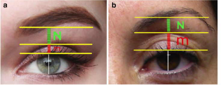
Note that both m and N are determined from the mid-pupillary line. As we age, m constantly becomes wider than N, causing the eye to sink in.
The goal of any rejuvenation procedure should be to restore the correct ratio of m and N (m/N = 1/2) with an optimal value of m 3–4 mm.
There are three groups of the degree of overhang of the upper eyelid:
* Group I — a congenital condition that causes the eye to look sunken. In this case, the N exponent is very short, and m is always wider than N. The main characteristic of the sunken eye — the presence of a clear separation between the visible area of the skin, the pretarsal part, and the orbital part of the eyelid, due to the deficiency of the preaponeurotic fat package, which undergoes progressive depletion against the background of bone thinning.
Read also: Chemical blepharoplasty: correction of the periorbital area with peeling
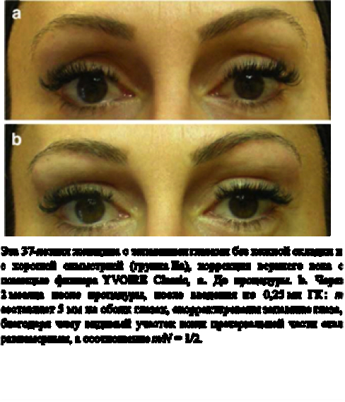
- Group II includes sunken eyes of the following types: (IIa) sunken eyes without a skin fold; (IIb) sunken eyes with moderate skin fold; (IIc) sunken eyes with pronounced skin fold; (IId) sunken eyes with dermatochalasis. In all these cases, m ≥ N. It should note that groups I and IIa are similar, but in the former case it is a congenital condition.
Read also: Technique for effectively removing forehead wrinkles
* Group III includes the so-called "complete" eyes. Two different cases are presented here: "complete"; eye with moderate skin fold (IIIa) and "full" eye with dermatochalasis (IIIb). In group IIIa, the ratio between m and N is not disturbed, but the volume of the orbital part of the eyelid is insufficient. In group IIIb, there is a sufficient volume of the visible area of the skin of the preseptal part, but due to pronounced dermatochalasis, m becomes less than 1 mm.
Clinical trials of upper eyelid correction methods
In a clinical study involving more than 500 patients, YVOIRE Classic filler based on HA of the Academy of Scientific Beauty company was used to restore the loss of volume of the upper eyelid.
YVOIRE Classic filler is used for groups I, IIa, IIb, IIc, IIIa, since in all these cases, excess skin is either absent or occurs due to volume loss, and after restoration of the lost volume of the upper eyelid with the filler excess skin disappears.
In groups IId and IIIb, treatment should be by blepharoplasty, and only in group IId, surgery is accompanied by the use of YVOIRE Classic filler at least 2 months after blepharoplasty. During surgery, resection of the excess skin flap is sometimes associated with resection of the medial portion of the nasal fat pad and minimal resection of one.
Rules for the prevention of side effects of facial skin peeling
Outcomes were assessed based on patient satisfaction and at least 1 year of clinical follow-up. The analysis of photographs taken before and after treatment was also taken into account. A total of 447 patients were women and 53 — men. In all cases, using the YVOIRE Classic filler, the correct ratio of m and N was restored, which indicates a high clinical efficacy of the drug and the duration of the result, which eliminates the need to repeat the procedure throughout the year.
Also, when comparing the results of correction of the periorbital region in patients who needed blepharoplasty, it was found that YVOIRE Classic filler can be used alone or in combination with surgery.
Follow our news on Facebook!
The right strategy should be chosen on the basis of a clear classification. Upper eyelid contouring with YVOIRE Classic filler — safe, simple and effective method with long lasting results. The YVOIRE Classic upper eyelid correction technique can also be used to restore volume loss after blepharoplasty and eye asymmetry in young adults.
Based on clinical study data, the duration of effect of YVOIRE Classic filler by Academy of Scientific Beauty is quite impressive when compared to other areas of the face. The longest follow-up period was 4 years, assuming the initial volume was maintained.
More useful information on our YouTube-channel:
Share:


Add a comment