

Dermal fillers for facial soft tissue augmentation are generally considered safe. Adverse events that occur after the administration of such drugs are in most cases mild and temporary. At the same time, the cosmetologist should not forget about serious complications that can develop after filling the nasolabial folds, nasolacrimal sulcus and other areas of the face. They are associated with circulatory disorders caused by occlusion and / or compression of the vessel by the filler. Damage to the vessels after a couple of hours leads to reticulation, and within a few days can provoke total skin necrosis.
Some patients do not have the typical symptoms of injection disorders, as in the case presented by Dr. Patrick Treacy in the article estet-portal.com.
- Case description: vein occlusion after filling of nasolabial folds with hyaluronic acid
- Management of vein occlusion and tissue necrosis after HA injections
Case description: vein occlusion after filling of nasolabial folds with hyaluronic acid
A 34-year-old female patient underwent correction of the nasolabial fold and marionette lines on the left side of her face. The procedure was uneventful, however on the second day after injections the woman went to the clinic complaining of the appearance of blue-black area of skin in the nasolabial and malar area. Due to the color of the affected area, the nurse mistook the injury for hematoma and prescribed arnica to the patient.
On the fourth day after the filling of the nasolabial fold, a bubble formed on the affected area, rejection of necrotic masses began. The patient went to the therapist, who, assuming the presence of an infection, prescribed antibiotic therapy.

Fig. 1: patient on the seventh day after HA injections
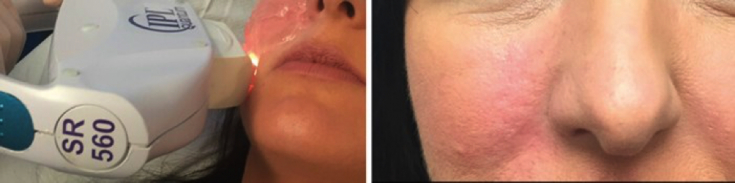
Fig. 2: patient on day 61 after IPL therapy
On the seventh day the patient's condition worsened. The woman went to the emergency room, who cleaned the wound and took no further action.
Nine days after the injections, the patient went to Dr. Tracy, who diagnosed vascular damage with the following:
- destruction of the skin;
- demarcation;
- ulcerations;
- rejection of necrotic masses.
Subscribe to our channel in Telegram!
The doctor believes that occlusion of the vein, and not direct blockage of the artery with the HA filler, was the cause of the complications, due to:
- late onset of symptoms (on the second or third day after injection);
- absence of pain;
- preservation of the normal color of the skin during the procedure (no blanching of the skin was observed).
Because the "golden" the recovery period was missed, the doctor developed the HELPIR technique specifically to work with this case.
Management of vein occlusion and tissue necrosis after HA injections
The HELPIR method that Dr. Tracy developed for the management of vein occlusion with subsequent tissue necrosis involves the use of:
H = hyperbaric oxygen
E = epithelial stimulation
L = low intensity laser therapy (633 nm)
P = platelet-rich plasma
I = IPL therapy
R = laser resurfacing
Having ruled out allergic reactions to hyaluronidase, the patient was immediately injected with 750 IU. Hyalase (hyaluronidase) in a dilute solution of 2% lidocaine to increase vasodilation and reduce the burning sensation caused by the enzyme.
The patient started taking 20 mg of Cialis (tadalafil), which improves blood flow due to its vasodilating effect.
Dr. Tracy prefers oral nitrates as they work for 48 hours, including at night, as opposed to Nitropaste, which must be applied every hour.
The patient was given 100mg of cortisone intravenously and 4mg of dexamethasone orally in case of vein occlusion due to edema and to reduce the inflammation caused by necrosis.
In order to prevent infections, the patient continued to take antibiotics and received daily hyperbaric oxygen therapy (the pressure was gradually increased from 75% to a maximum).
Inducing re-epithelialization by PRP and low-intensity laser therapy
Platelet-rich plasma promotes tissue regeneration and is becoming a valuable method for promoting wound healing in aesthetic medicine. From the patient's blood, PRP is obtained by centrifugation. The drug is saturated with growth factors, which trigger the mechanisms of tissue repair. These include:
- platelet growth factor (PDGF);
- transforming growth factor beta (TGF-b) 1 and 2;
- vascular endothelial growth factor (VEGF).
All of them are involved in the angiogenic cascade, which accelerates the recovery of damage to hard and soft tissues. The author foresaw that the VEGF factor would provoke excessive vascularization of the problem area, which would need to be treated with a vascular laser.
Low Level Laser Therapy (LLLT) – rapidly advancing technology stimulating:
- wound healing;
- reduction of pain and inflammation;
- restore tissue function.
Photons are absorbed by mitochondrial chromophores in skin cells. As a result, electron transport is carried out, nitric oxide ATP is released, blood flow increases, the number of reactive oxygen species increases, and various signaling pathways are activated. Stem cell activation promotes enhanced skin regeneration. To eliminate the consequences of vein occlusion in the clinic, the author used the Omnilux 633 laser, and for home use, the patient was given a portable device Genosys Omega.
Read also: Complications of contour plastics: when «beauty injections» not good
Hyperbaric oxygenation increases the oxygen gradient between the center and periphery of the wound, providing a powerful angiogenic stimulus. Along with the proliferation of fibroblasts, neovascularization increases.
To avoid scarring, it is important to restore the epithelium in the affected area as quickly as possible.
Hyperoxia in normal tissues causes vasoconstriction, which reduces post-traumatic tissue edema, contributing to the healing of necrosis. However, this vasoconstriction does not lead to hypoxia, as it is overcompensated by increased plasma oxygen levels as well as microvascular blood flow.
Hyperbaric oxygen therapy increases the production of free radicals, which oxidize proteins and membrane lipids, damage DNA and inhibit the metabolic functions of bacteria.
Read also: First aid for central retinal artery occlusion after filler injections
Oxygen is required for the hydroxylation of lysine and proline residues during collagen synthesis, as well as for collagen cross-linking and maturation necessary for wound healing. The lack of oxygen is corrected during hyperbaric oxygen therapy, which leads to the formation of the required amount of mature collagen.
Fibroblast proliferation and neocollagenesis are triggered by low-intensity laser therapy and platelet-rich plasma growth factors. This leads to unwanted angiogenesis, which is further corrected by IPL therapy.
Adapted from The PMFA Journal.
Subscribe to our YouTube-channel!
Share:


Add a comment