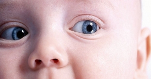
At the birth of a child, the structure and function of some of its organs differ significantly from those of adults over a period of time. So, the visual analyzer in a child after birth is rebuilt to new living conditions and gradually develops, acquiring the outlines of an adult eye only by the age of 10-12. What changes in the visual analyzer are physiological?
How should the visual analyzer differ in children?
It is important to know the periods of formation of all components of the visual analyzer for the timely detection of congenital or acquired pathologies of the eye in a child. The slit of the child's eye is narrow and corresponds to the size of the pupil. The frequency of blinking the eyes of a child is 2-3 times per minute, which is 7 times less than in adults. The eyelids during sleep often do not close completely, a blue stripe of the sclera is visible. By the age of 2, the frequency of blinking increases to 7-8 times per minute.
The lacrimal gland does not begin to function until a few weeks after birth, so in the first month of life, the baby cries without tears.
The internal components of the visual analyzer in children also have some peculiarities. These include:
- orbit;
- conjunctiva;
- cornea;
- sclera;
- front camera;
- iris;
- ciliary body;
- fundus.
Features of the visual analyzer in newborns. How to distinguish pathology?
The orbit in children under one year old is relatively small, which makes the eyes visually look larger. The horizontal size of the eye sockets is always greater than the vertical size, but the convergence of the axes of the eye sockets, on the contrary, is less. This sometimes resembles a clinic of convergent strabismus. The temporal wings of the sphenoid bones are underdeveloped at birth, so the palpebral fissures in children are wide. Tooth germs are close to the contents of the orbit, which increases the likelihood of odontogenic infection. The development of the eye socket of the visual analyzer ends by the age of 7.
The conjunctiva of a newborn is very thin, not moist enough, has low sensitivity and can be easily injured. Already by the age of 3 months, it becomes more sensitive and moist. Pronounced moisture and the presence of drawings on the conjunctiva indicate an inflammatory process or congenital glaucoma.
What is the reason for the frequent ingress of foreign bodies into the visual analyzer of children?
The cornea of the optic analyzer may be dull after birth. Already after the first week of life, it becomes transparent. It is important to distinguish physiological dullness from corneal edema in congenital glaucoma. Increased pressure in glaucoma is removed by instillation of a hypertonic solution of 5% glucose. The first sign of congenital glaucoma in children is an increase in the diameter of the cornea. The normal diameter of the cornea is 9-9.5 mm. By the first year, it increases by 1 mm. The sensitivity of the cornea is very reduced in children up to 3 months. Weakened corneal reflex contributes to the entry of foreign bodies into the eyes, so it is important to frequently examine the child's eyes.

The sclera of the visual analyzer is thin, has a bluish tint, which disappears by 3 – summer age. But this should also be taken seriously, as blue sclera can be a sign of stretching due to high intracranial pressure.
The anterior chamber is small, has an acute angle. The root of the iris is gray-black, which is explained by the remnants of embryonic tissue, which is completely absorbed during the first year of life.
Features of the reaction of the pupils and the fundus of the visual apparatus of children
The iris has a bluish-gray color due to a small amount of pigment in it. Individual eye coloration begins to acquire by the end of the first year of life, the final establishment of eye color lasts up to 12 years. In one-year-old children, the direct and friendly reaction of the pupil is not clearly expressed, the pupils are poorly dilated with medications.
The ciliary body is in a state of spasm for six months after birth. This contributes to the development of myopic clinical refraction without cycloplegia, as well as the presence of hyperopic refraction after instillations of 1% homatropin.
The fundus of the visual analyzer in newborns has a pale pink color. The vascular network is clearly visible, the optic disc is pale, has a bluish-gray tint. This fact can be mistaken for optic disc atrophy.
Knowledge of the physiological characteristics of the visual analyzer in children is important for the detection of congenital pathologies.


Add a comment