

Features of the anatomical structure of the subcutaneous cervical muscle determine its influence on the aging process of the face. Recent foreign studies in the field of the anatomy of the subcutaneous muscles allow a different look at the methods of correcting age-related changes in the area of platysma, as well as the aging processes of the skin and some structures that help keep the muscles of the face in a taut state.
Alan Fogli:
- Professor of the International Association of Aesthetic Plastic Surgeons ISAPS;
- Honorary President of the National Organization for Plastic, Reconstructive and Aesthetic Surgery;
- Member of the French Association of Plastic, Reconstructive and Aesthetic Surgeons;
- Member of the Argentine Association (Buenos Aires) for Plastic, Reconstructive and Aesthetic Surgery;
- founder of Clinique de chirurgie esthetique du docteur Fogli Alain, Marseille (France).
Readers estet-portal.com offers a summary of the article by Professor Alan Fogli (France) about his own view on the methods of surgical correction of age-related changes in the muscles of the neck.
Subcutaneous cervical muscle and its role in the process of facial aging
The subcutaneous cervical muscle of a young man is a muscular lobe and forms an almost right angle with the horizontal part, it is located at the level of the rhizorius and almost fills the space below the collarbone (Fig. 1).
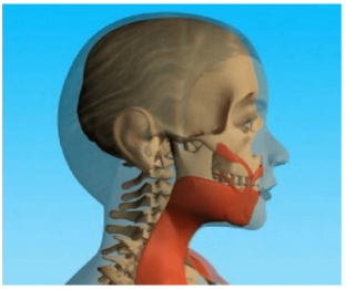
Fig. 1 Location of the platysma muscle in a young man
The upper fibers of the muscle are at first horizontal, then they bend in front of the earlobe, intersect at the lower jaw and become almost vertical, covering the anterolateral part of the neck. The fibers of the anterior part of the neck muscle are separated on both sides from the midline (Fig. 2).
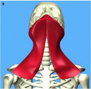
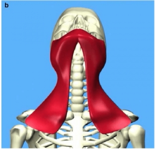
Fig. 2(a). Separation of the platysma muscle in a young person, (b) Association of the platysma muscle in an older person
According to studies conducted in the 1980s, the subcutaneous cervical muscles undergo the same changes in position during the aging process of the face. These studies were carried out in the anatomical laboratories of Prof. José Santini in Nice (France), Philippe Keix in Bordeaux (France) and Daniel Eglof in Lausanne (Switzerland).
The upper fibers of the platysma muscles are located on average 2-3 cm below the horizontal line at the rhizorius, and in young people they cross the lower jaw in front, while the fibers of the posterior edge of the muscle do not, or only slightly, change in aging process (Fig.3a and b).
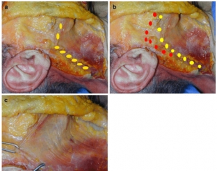
Fig. 3. (a) Position of the upper and lower edges of the platysma muscle in old age (right symbols), (b) Position of the upper and lower edges of the platysma muscle in old age (left symbols). Please note that, on the one hand, the platysma fibers move down and forward, and on the other hand, the posterior edge does not change during the aging process of the face. (c) Movement of the platysma muscle along a vertical vector (observed in young people.
The anterior margins of all the platysma muscles move anteriorly towards the margin of the subcutaneous cervical muscle, and their connection in the midline has already been studied by Connell and Gaol. It must be remembered that in young people they are located in front, and their condition depends on the severity of the platysma ligaments, which may be more or less visible, depending on the formation of the angle between the neck and chin (Fig. 2b).
The shape of the ligaments of platysma is not the result of muscle stretch and relaxation, but rather its sliding in the outer aponeurotic plane, which is enhanced by contraction. This process perfectly matches the logic of general muscle aging.
The role of skin condition in the process of facial aging
The ligaments between the skin and the platysma muscle are very tight, so there is no noticeable sliding between these tissues. Indeed, the platysma muscle has the same embryological origin as the skin. They undergo the aging process at the same time, due to which their close interaction is maintained.
Fixation structures to help correct the aging process of the face
Just like the retaining ligaments, some structures are not subject to repositioning as the face ages. These structures include the zygomatic periosteum, the parotid aponeurosis and Loret's fascia, which was the subject of an anatomical study by Labbe et al. This fascia was described by Loret as a landmark in parotid surgery when reaching the facial nerve trunk. Indeed, the subcutaneous ligaments anterior to the earlobe, as described by Furnas, are a dense white fibrous structure associated with the parotid fascia (Fig. 4a).
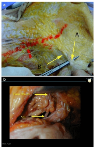
Fig. 4. (a) Ear platysma ligament (A) and prelobar fibrous tissue (B). (b) Superficial to deep tissues: the top arrow indicates the attachment plane, which is usually 0.7-1 cm below the skin surface; the lower arrow points to the trunk of the facial sockeye salmon, which is located at a depth of 2.5 cm under the surface of the skin
These various structures fuse together to form a fibrous, homogeneous, dense, and hard tissue that serves as an attachment for the platysma fibers. They are located in incisions 0.7 to 1 cm below the skin surface, while the facial nerve trunk is 2.5 cm below the skin surface (Fig. 4b). Thus, there is no danger of the platysma becoming tightly anchored at the level of this fibrous structure.
Surgical findings regarding the aging process of the face
The surgical findings from these anatomical findings are extremely significant, as they do not correspond to the evolution of surgical principles that have been studied and practiced for decades, but they follow a logic that is completely opposite in relation to earlier knowledge for the following reasons.
Firstly, the separation of skin and muscle is not only useless and illogical, but also reduces the effectiveness of the surgical intervention. The connection between skin and dermal muscle changes slightly with aging. It is a very large adjacent attachment surface and an amalgamation of numerous microstructures that hold the skin together. In addition to skin devitalization, their separation reduces the effectiveness of invasive techniques. The tissues fit together on the aponeurotic superficial plane.
The first surgical conclusion is not to separate them, but to create a muscular support that will allow them to be lifted together and thereby restore the anatomy as it exists in young people (Fig. 3c). The branch is especially illogical and because it damages the posterior margin of the platysma, which does not undergo changes with aging.
Furthermore, separation of the posterior edge results in stretching and fixation of the muscle at the back, in a position that is unnatural for both old and young people. There is no reliable structure for posterior anchoring. Fibrous tissues always move forward under pressure and give the appearance of sagging. This creates an anatomical structure that explains the low efficiency and unnatural aspect of some of the results.
Secondly, the suture between the muscles of the midline platysma, which is formed in the submental approach, is also counterintuitive in this concept, because it separates the dermal muscles in an anatomical position characteristic of senile patients. This structure should not form if the platysma muscles have been significantly raised and tightly secured to the anchoring structures mentioned earlier.
Alan Fogli will present his own method of correcting age-related changes in the area of platysma and the effect on the aging process of the skin and some structures that allow keeping facial muscles in a taut state at the ISAPS World Congress, which will be held in Kyiv on May 19-20 !
In the following article, readers of estet-portal.com are offered a description of Alan Fogli's special surgical technique, based on the movement of facial tissues and platysma fibers to the places where they are in young people.
Share:


Add a comment