

The first signs of skin aging appear, as a rule, in the periorbital zone. Patients of cosmetology clinics are convinced that the area around the eyes – key in aesthetic terms, it largely determines the image of a young face. Therefore, procedures for the rejuvenation of the periorbital zone are among the most popular. To correct the area around the eyes, surgical blepharoplasty and hardware laser exposure are most often used. However, these methods have many limitations and side effects. Therefore, as estet-portal.com notes, more and more patients are interested in minimally invasive techniques – e.g. plasma excision.
What are the features of different methods of correction of the area around the eyes
Surgical blepharoplasty is considered the most effective method for correcting age-related changes in the upper and lower eyelids. With a sufficiently high professional qualification of the surgeon, it gives excellent results, such an operation is not performed with some contraindications associated with the internal diseases of the patient. In addition, after such a surgical intervention, although rarely, complications occur – for example, ectropion, lagophthalmos, ptosis, dry eye syndrome, scarring, and some others.
Laser procedures for the correction of the periorbital zone have a much smaller list of restrictions and complications than blepharoplasty. However, to protect the eyes from laser exposure, the doctor uses special devices that are very inconvenient for patients. Besides,
The choice of a correction technique for the area around the eyes is up to the doctor, who assesses the patient's condition, possible risks of intervention and patient expectations. Plasma excision with Plexr,
The advantages of plasma excision in cosmetic correction of the eye area
Plasma excision – it is a promising, fast and safe minimally invasive solution to the problem of age-related changes in the eye area. Its success is due to its mechanism of action: energy from the device ionizes the gases in the air between the handpiece and the skin and creates a plasma that is transferred to the superficial layer of the skin.
Compared to the monopolar radioscalpel, the main advantage of the plasma excision technique is that no electrical current is transmitted through the patient's body.
Thanks to this, even patients with pacemakers in the heart can be corrected. Compared with laser correction of the periorbital zone, the plasma technique acts only in the surface layer of the skin, without contact with the basement membrane, so the risk of visual impairment or eye injury after plasma excision is minimal.
Prior to the procedure, a simple anesthetic cream is applied to the area to be treated, and no additional eye protection is required as the patient's vision is not compromised. After all, the generated heat is absorbed only by the treated area of the skin and is not transferred to the surrounding tissues or subcutaneous tissue.The main advantages of this technique are:• minimum contraindications,
• low pain during surgery, • speed of the procedure,• fast recovery,
• excellent value for money.
 Thus, the use of Plexr for the correction of age-related changes in the area around the eyes is appropriate.
Clinical studies on the effectiveness of plasma excision with the Plexr
Ten patients underwent 3 plasma excision procedures. Photographs and images were obtained at baseline and 4-6 weeks after the final plasma excision procedure. Patient inclusion criteria were as follows: skin phototype I-III and the presence of moderate or severe laxity of the upper eyelid crease according to the facial laxity rating scale. Exclusion criteria were the following: active infection, tendency to form hypertrophic scars, previous medical procedures in this area (Botox injections, fillers, chemical peels, laser treatment and intense pulsed radiation) or use of oral retinoids within the last 6 months. All patients provided written consent.
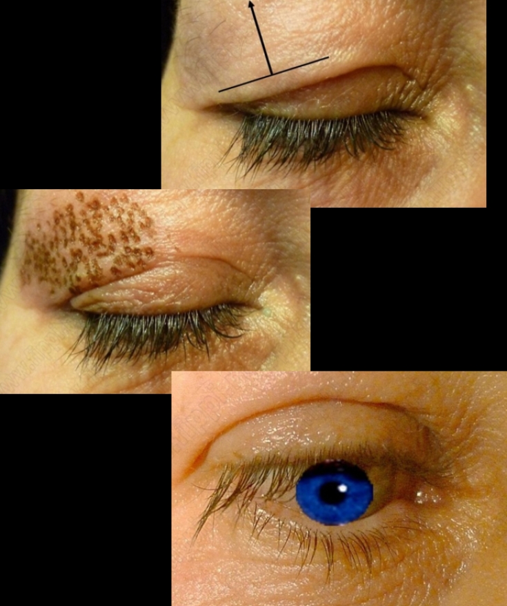
After gentle cleansing of the respective tissue area, an anesthetic cream was applied and left for 30 minutes. The area was then cleaned and disinfected again. All patients underwent the procedure using the Plexr instrument. The device was set to the processing mode of a single section (point by point) and one point was processed for no longer than 2 seconds. The points were located in close proximity to each other and did not intersect.
Plasma excision was performed on an excess skin flap during three sessions with an interval of 30 days. At each session, an area equal to 30% of the surface of the upper eyelid was treated. The lateral areas of the eyelid were treated in the first session, in the second they moved to the central area, and the medial surface was treated in the third (final) session.The area was then cleaned and disinfected again. All patients underwent the procedure using the Plexr instrument. The device was set to the processing mode of a single section (point by point) and one point was processed for no longer than 2 seconds. The points were located in close proximity to each other and did not intersect.
Plasma excision was performed on an excess skin flap during three sessions with an interval of 30 days. At each session, an area equal to 30% of the surface of the upper eyelid was treated. The lateral areas of the eyelid were treated in the first session, in the second they moved to the central area, and the medial surface was treated in the third (final) session. The area was then cleaned and disinfected again. All patients underwent the procedure using the Plexr instrument. The device was set to the processing mode of a single section (point by point) and one point was processed for no longer than 2 seconds. The points were located in close proximity to each other and did not intersect.
Plasma excision was performed on an excess skin flap during three sessions with an interval of 30 days. At each session, an area equal to 30% of the surface of the upper eyelid was treated. The lateral areas of the eyelid were treated in the first session, in the second they moved to the central area, and the medial surface was treated in the third (final) session.
The device was set to the processing mode of a single section (point by point) and one point was processed for no longer than 2 seconds. The points were located in close proximity to each other and did not intersect.
The device was set to the processing mode of a single section (point by point) and one point was processed for no longer than 2 seconds. The points were located in close proximity to each other and did not intersect.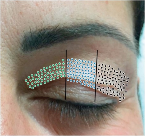
An example of treatment of upper eyelid areas: black dots – 1st session, blue – 2nd session, green – 3rd sea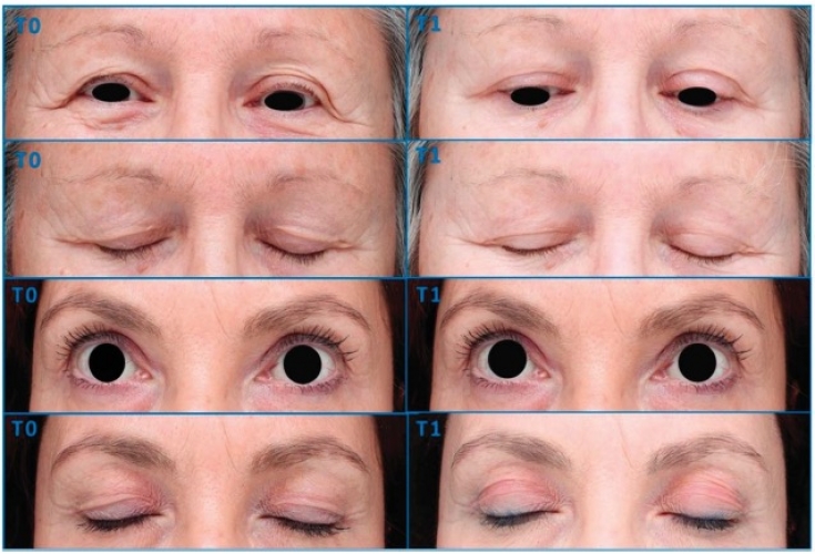
Examples of clinical improvement after 3 plasma excision sessions
Using reflective confocal microscopy, it was found that the positive clinical results of correction of the area around the eyes by plasma excision are associated with the reorganization and remodeling of collagen. New thick shiny collagen fibers with a parallel orientation were observed, which provided skin tightening and eliminated the sagging of the eyelid without tissue excision.
Thus, the plasma excision technique using the Plexr apparatus of the Italian company GMV SRL, which is provided by
Share:


Add a comment