
In aesthetic surgery, correction of the periobitual area is one of the most requested surgical procedures. Age-related changes in the face begin to appear quite early in this zone, and in the modern world, people want to preserve their beauty and youth for as long as possible. Surgical interventions in the periorbital region require high qualification and extensive experience of a plastic surgeon, in addition, it is very important to properly prepare for such an operation. Knowledge of the anatomical features of the structure, as well as the basics of surgical rejuvenation of the periorbital region and eyelids, allows the surgeon to satisfy all the wishes of the patient as much as possible.
Preparation for surgical intervention in the periorbital region
In modern aesthetic surgery of the face, the periorbital region is distinguished as a complex of anatomical elements mutually influencing each other, which undergo gravitational changes in a certain sequence. The composition of the periorbital region includes the eyebrows, the glabella and the base of the nasal pyramid, the upper and lower eyelids, the corners of the eyes, as well as the tissues located outward from the lateral edges of the orbits, the so-called "crow's feet" area. At a young age, the orbit has the shape of an ellipse, which is somewhat inclined in accordance with the projection of the corners of the eyes. As the ellipse ages, under the influence of gravity, turns into a circle, which is important to consider when planning the correction of the periorbital region.
Periorbital area:
- Planning surgery to correct the periorbital area;
- Selection of the volume of surgery for the correction of the periorbital region and eyelids;
- structural features of the upper and lower eyelids.
Planning surgery for correction of the periorbital region
In planning surgery for the correction of the periorbital zone of the face, several of the most important points of the preoperative examination of the patient can be distinguished:
- determination of the patient's vision, functions of the lacrimal gland, conjunctiva and lacrimal ducts;
- evaluation of the position and shape of the eyebrows, the severity and location of glabella wrinkles, the ratio of the height of the lateral thirds to their medial parts;
- specifying the degree of protrusion of the eyeball in relation to the lower edge of the orbit;
- assessment of the characteristics of the layers of the upper and lower eyelids, as well as the levels of the position of their ciliary edges;
- examination of the position and stability of the lateral canthal ligaments;
- determination of the severity of age-related changes in the mid-medial part of the face, the position of the eyelid-buccal and lacrimal grooves in relation to the lower orbital edge.
Selection of the volume of surgery for the correction of the periorbital region and eyelids
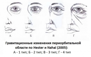
In accordance with the classification of age-related changes in the periorbital zone according to Hester and Nahai, with type 1, only the initial signs of the appearance of aging are determined, while with type 4, their maximum severity is observed. Thus, performing isolated upper and lower blepharoplasty is justified when all age-related changes are concentrated only within the orbit, that is, in types 1 and 2. Surgical correction of age-related changes in the periorbital zone in patients of groups 3 and 4 cannot be limited only to blepharoplasty. In these cases, the operation should include intervention in certain related areas.
Features of the structure of the upper and lower eyelids
To perform surgical correction of the periorbital region and eyelids, it is fundamentally important to take into account the structural features of the upper and lower eyelids. To clarify the characteristics of the layers of the upper eyelid, it is necessary to evaluate the following parameters:
- front lamella – clarify the presence of true dermachalasis of the eyelid, the location of the tarsal fold, the state of the orbicular muscle of the eye;
- medium lamella – characterize upper fatty hernias;
- rear lamella – assess the level of the position of the ciliary edge of the upper eyelid in relation to the pupil and the upper edge of the iris.
In the lower eyelid, it is important to assess the condition of the following layers:
- anterior lamella, which includes the skin and orbicular muscle of the eye;
- middle lamella – namely, its retroseptal adipose tissue and intraorbital fascial septum;
- rear lamella – supporting ligamentous structures of the lower eyelid, conjunctiva and Müller's inferior muscle.
Share:


Add a comment