


"You can only cure what you can see" — these are the words of Harry Keir, one of the founders of the field of dental microscopy. He's right. Often, medical errors are due precisely to the fact that the specialist does not have the necessary equipment for the correct diagnosis of diseases and the quality control of treatment. The dental microscope solves these problems. Thanks to modern dental optics, it is possible to achieve a multiple increase in images when examining the condition of the teeth, to treat them with high quality.
- What is a Dental Microscope
- Comfort during dental treatment — important question
- Root Canal Revision: Tooth Rescue Instead of Extraction
- Highlights on the use of microscopes in dentistry
What is a dental microscope
Magnifying devices are used in many areas of medicine — in ophthalmology, neurosurgery, vascular surgery. Since the 80s of the last century their began to be used in stomatology. Dental technique gives an image magnification from 3 to 40 times, therefore it is indispensable in the work of endodontists.
Models of dental microscopes are diverse in dimensions, technical characteristics and installation methods, so each clinic has the opportunity to choose the right equipment, taking into account the specifics of the work of doctors — therapists, surgeons, endodontists.
Dental microscope is most often used for the following purposes:
-
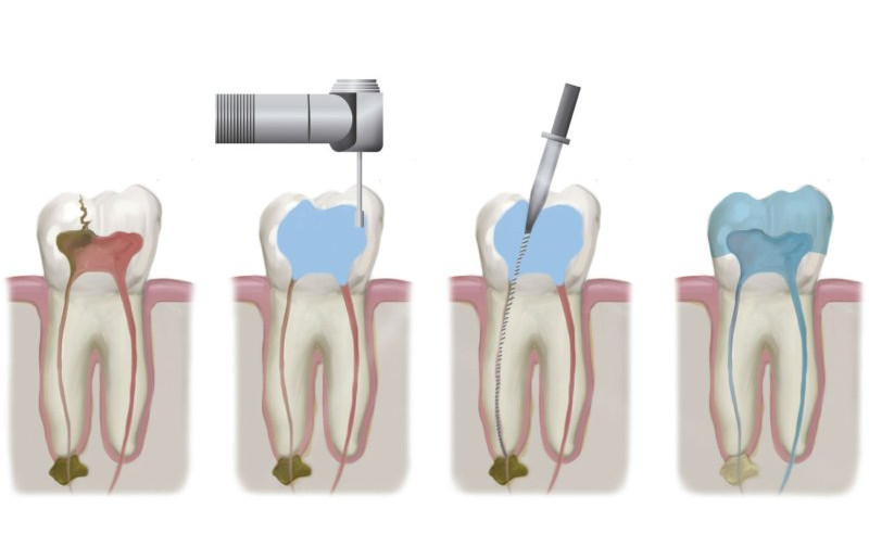
Diagnosis of the state of root canals. Their diameter — 1-3 mm. Without a microscope, the doctor has to diagnose literally blindly, which is fraught with errors.
-
Extraction of foreign objects. Often, in the process of dental treatment, particles of filling materials get into the canals. This leads to inflammation.
-
Removal of pieces of instruments. Even if dentists use instruments made of heavy-duty materials, they may break due to their thinness. Thus, metal fragments remain in the channels, which patients do not suspect.
-
Revision (retreatment) of root canals. If the tooth was treated poorly, serious problems arise. The canals must be retreated, otherwise the tooth will have to be removed.
Thanks to detailed visualization with the help of a good microscope, the doctor can correctly diagnose diseases, correct mistakes made by specialists who worked before. And for the patient, this is an opportunity to save teeth that would otherwise have to be removed.

Read also: Proper care braces: features of oral hygiene
Comfort during dental treatment — important question
Effective painkillers have long been available, but the fear of dentists hasn decreased. One of the reasons why we are afraid to treat our teeth — discomfort. The patient has to sit in an uncomfortable position, change from one painful position to another. The neck is numb, and you can’t move. The most unpleasant thing is that the doctor has to suffer in the same way in order to examine the teeth in detail, and this ends with pain in the back.
If the examination and treatment is carried out using a dental microscope, the problem of discomfort for the dentist and patient is resolved automatically. The technique is designed so that the doctor himself regulates the position of the equipment. He does not have to bend at unthinkable angles and turn the patient's head. Everyone wins, feels comfortable and calm.

Read also: How to treat dental caries: all about diagnosis and prevention
Root canal revision: saving the tooth instead of extracting it
Until the introduction of microscopes in dental clinics, re-treatment of teeth rarely gave the expected results. Most often, poorly treated canals became inflamed, which could lead to atrophy of the bone area, tissue necrosis near the site of infection. Dentists honestly carried out an audit of the canals, but the treatment was not always successful. After a long torment, the teeth were removed. Dental optics has radically changed the situation.
With a multiple increase, the doctor can examine in detail even the most complex channels, thin branches, determine the presence of cavities or foreign objects. This greatly increases the chances of saving the tooth. For retreatment, you will have to visit the dentist’s office at least twice, but it’s still much better and cheaper than removing a tooth, and then prosthetics.
Read also: Tooth abscess: how to avoid acute purulent inflammation
Highlights on the use of microscopes in dentistry
Most large clinics have long been equipped with high-quality dental equipment. A dental microscope is a must, because it helps to solve many problems:
-
Examine and fill root canals. They can be complex, tortuous, or overly thin. With using a microscope, you can determine the configuration and features of the canals, treat them qualitatively.
-
Root canals are retreated. If the clinic is equipped with good optics, the chances of saving a tooth that was previously treated unsuccessfully increase.
-
Find and remove foreign bodies.In canals often remain pieces of filling materials, fragments of instruments. The patient does not know about them, but suffers from inflammations.
When choosing a doctor, be sure to take an interest in the technical equipment of his office, especially the presence of a microscope, because your dental health depends on it.
Read also: Why do gums bleed and what to do about it
You might be interested in: How to place a filling.
Add a comment