
The problem of ovarian endometriosis has affected millions of women on the planet today. It is this type of endometriosis that causes particular concern for the category of such patients, since ovarian endometriosis in some cases tends to become malignant and form malignant ovarian tumors.
Endometrioid ovarian cysts have a characteristic clinical picture and require the earliest possible detection and administration of an effective treatment regimen in order to prevent oncological pathology and restore the normal function of the woman's reproductive system.
Ovarian endometriosis: main symptoms and diagnostic methods
Endometriosis of the ovaries occurs as a result of lymphogenous or hematogenous entry of endometrial cells into the region of the uterine appendages. Often, ovarian endometriosis is manifested by the formation of cysts and pseudocysts, which give a very characteristic clinical picture.
The symptoms of endometrioid cysts significantly impair a woman's quality of life, affect her general condition and disrupt reproductive function. In addition, endometrioid cysts in some cases can transform into malignant ovarian tumors. The characteristic clinical picture and modern diagnostic methods help in the detection of ovarian endometriosis.
Ovarian endometriosis:
- main forms of ovarian endometriosis depending on their structure;
- characteristic clinical picture of ovarian endometriosis;
- Basic methods for diagnosing ovarian endometriosis.
The main forms of ovarian endometriosis depending on their structure
Ovarian endometriosis is manifested by the formation of pseudocysts up to one centimeter in diameter, which are filled with a brown mass. When such pseudocysts merge, endometriomas or the so-called "chocolate cysts" are formed. Endometrioid lesions are localized most often in the cortical layer of the ovaries, but sometimes they can also affect the medulla. Depending on the structure, the following forms of ovarian endometriosis are distinguished:
- glandular endometriosis;
- cystic endometriosis;
- glandular cystic endometriosis;
- ovarian stromal endometriosis.
It is the glandular-cystic form of ovarian endometriosis that is most often prone to excessive proliferation and malignant transformation. Endometrioid pseudocysts are lined with connective tissue, and endometriomas – columnar or cuboidal epithelium. The dark brown color of the contents of these cysts is due to the large amount of hemosiderin in the cystic mass.
Characteristic clinical picture of ovarian endometriosis
In the presence of small foci of ovarian endometriosis, the clinical picture is absent in most cases. The first symptoms of the disease occur when pseudocysts coalesce and form endometriomas. In this case, the parietal and visceral peritoneum is drawn into the pathological process, and as a result of the subsequent spread of pathological foci, adhesive processes are formed.
At the same time, the patient feels constant pain in the lower abdomen of a dull, aching nature, which can radiate to the rectum and perineum. During menstruation, the pain increases significantly. Due to the adhesive process, pain syndrome also occurs during physical exertion and sexual intercourse. Patients with ovarian endometriosis are also characterized by menstrual dysfunction by the type of algomenorrhea.
Basic methods for diagnosing ovarian endometriosis
Diagnosis of ovarian endometriosis is based on anamnestic data, where the patient complains of chronic pain, as well as on the results of a gynecological examination and instrumental diagnostic methods.
Palpation is uninformative in the presence of small endometrioid pseudocysts, but endometriomas are palpated as stiff elastic formations localized posterior to the uterus, limiting its mobility. After menstruation, the size of endometriomas increases.
On ultrasound, the presence of pseudocysts can be indicated by hypoechoic inclusions on the surface of the ovaries and thickening of their albuginea as a result of the formation of adhesions.
Ultrasonographically, endometriomas are visualized as rounded formations with a pronounced echopositive capsule, localized behind the uterus, having a finely dispersed echopositive suspension against the background of liquid contents.
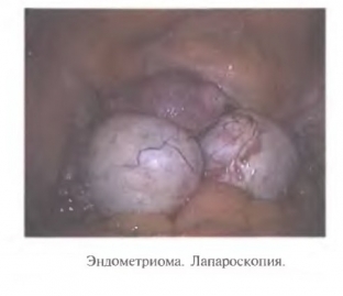
Laparoscopy allows not only to clarify the diagnosis of ovarian endometriosis, but also to directly treat this pathology.
Share:


Add a comment