
Surgical treatment of patients with such severe and pronounced eyelid deformities as neurofibromatosis of the periocular region presents significant difficulties, since in order to achieve a stable and functional result, it is necessary to radically remove the tumor, excise bone structures, and restore the anatomical structure of the affected eyelid. Read about how blepharoplasty is performed in case of neurofibromatosis of varying degrees, and what is necessary to achieve a good cosmetic result, read on estet-portal.com.
Clinical case
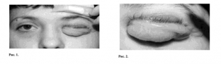
Three patients with type I neurofibromatosis applied to the eye microsurgery clinic – eyelid involvement was observed, the diagnosis was confirmed histologically and according to the clinical classification. So, the patients were diagnosed with:
- patient 1: grade 1 disease - blepharoneurofibromatosis with a large number of isolated soft tissue lesions in the periocular region,
- patient 2: disease 2.1 degree – neurofibromatosis with damage to soft tissues not only of the orbit, but also of the temporal region, with preservation of unchanged visual functions,
- Patient 3: Grade 2.2 disease - blepharoneurofibromatosis with soft tissue damage in the orbit and temple, with minimal bone damage, visual functions are preserved.
The second and third patients have previously been operated on in other specialized medical institutions.
Preparation for blepharoplasty
Taking into account the peculiarities of the course of the disease and the degree of damage to the eyelids, in preparation for surgical intervention, the patients underwent desensitizing therapy, all of them were prescribed vitamins A, E, group B.
When planning a surgical intervention and calculating blepharoplasty, the principle of maximum selection and similarity of replaced eyelid tissues with those that will be moved to replace them was observed.
Treatment: features of blepharoplasty for neurofibromatosis of varying degrees.
The amount of surgical intervention performed for each patient depended on the degree of eyelid involvement. In patient 1, during blepharoplasty, an incision was made parallel to the edge of the eyelid in accordance with the localization of the tumor. The tumor tissue was separated from the skin with the preservation of the flap for subsequent plasty. The tumor tissue was excised simultaneously with the surrounding tissues, the removal was performed with an indent of 5 mm from the edge of the tumor with excision to the cartilage or bone. The resulting eyelid defects were covered with displaced tissues, which were fixed to the underlying tissues. Interrupted sutures were placed through the fixed and underlying tissues.
In patients 2 and 3, during blepharoplasty surgery, a skin incision was made along the alleged skin fold on the upper eyelid. Next, the skin was cut off in the up and down directions, the affected tissues, including cartilaginous plates in the outer third of the eye, were completely removed. Cartilage was excised within the unchanged tissue of the orbit, retreating from the edge by 5 mm. After excision, thin eyelid skin and conjunctiva remained, so the eyelid frame was restored using an alloplant, which was sutured to the remaining cartilage from the inside of the eyelid. The excess thin stretched eyelid skin was partially resected, the remaining tissue of the eyelid and conjunctiva was moved and fixed to the underlying unchanged tissues with interrupted vicryl 6.0 sutures.
Results of blepharoplasty for neurofibromatosis
The results of blepharoplasty operations performed on all three patients were evaluated 6 and 12 months after surgery. Operations in the first and second patients were uneventful. In the third patient, who had the largest amount of surgical intervention, lymphostasis developed in the postoperative period, and due to prolonged edema, partial necrosis of the skin of the eyelid occurred. This required closing the defect by moving the skin flap from the side of the temple.
Read also: Modeling the fold of the eyelid: features of surgery
In the postoperative period, the patients received local and general, according to indications, anti-inflammatory treatment, trophic and resolving therapy was prescribed.
During the examination before discharge, the following results were recorded: the palpebral fissure recovered almost to the correct natural shape and acquired a natural size. The edge of the eyelid after blepharoplasty is somewhat thickened, eyelashes with irregular growth remain in places.
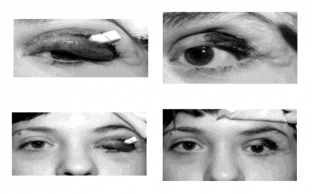
Functional and cosmetic results of the blepharoplasty were assessed as good in the first and second patients, in the third patient – as satisfactory. Within 1-2 years, all three operated patients noted a stable remission.
As a result of blepharoplasty surgery, it was possible to eliminate eyelid deformity due to neurofibromatosis and restore its functions.
Share:


Add a comment